. 2024 Oct 10:15:1469353.
doi: 10.3389/fimmu.2024.1469353. eCollection 2024.

Transition from acute kidney injury to chronic kidney disease in a long-term murine model of Shiga toxin-induced hemolytic-uremic syndrome

Affiliations

Transition from acute kidney injury to chronic kidney disease in a long-term murine model of Shiga toxin-induced hemolytic-uremic syndrome

Jamila Wegener et al. Front Immunol. .

Abstract

Introduction: Up to 40% of patients with typical hemolytic-uremic syndrome (HUS), characterized by microangiopathic hemolytic anemia and acute kidney injury (AKI), develop long-term consequences, most prominently chronic kidney disease (CKD). The transition from AKI to CKD, particularly in the context of HUS, is not yet fully understood. The objective of this study was to establish and characterize a Shiga toxin (Stx)-induced long-term HUS model to facilitate the study of mechanisms underlying the AKI-to-CKD transition.

Methods: C57BL/6J mice were subjected to 5, 10, 15, or 20 ng/kg Stx on days 0, 3, and 6 of the experiment and were sacrificed on day 14 or day 21 to identify the critical time of turnover from the acute to the chronic state of HUS disease.

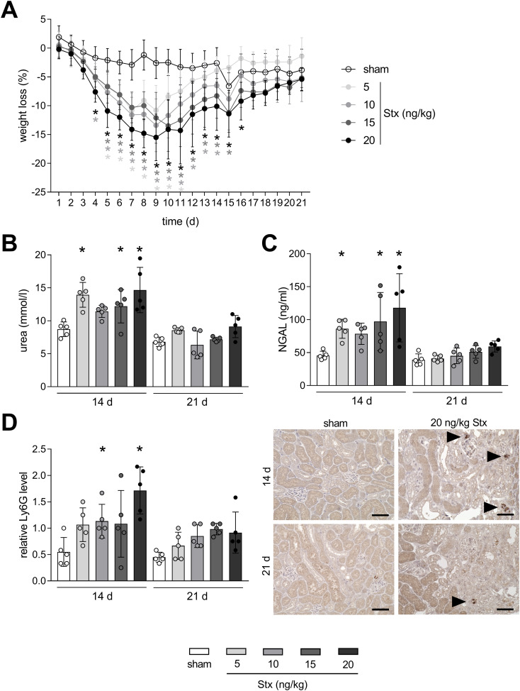
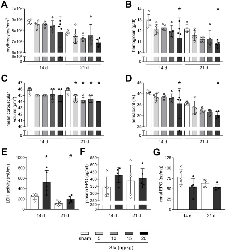
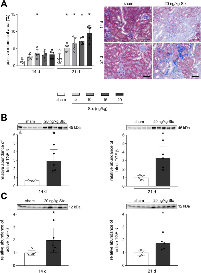
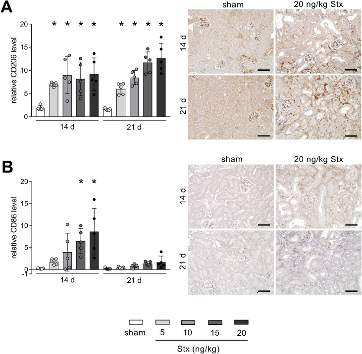
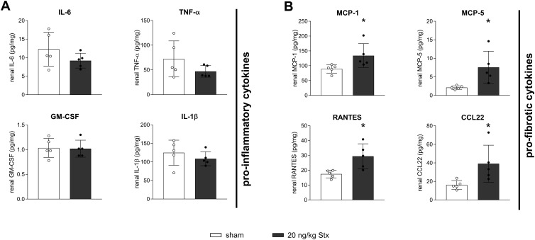
Results: Acute disease, indicated by weight loss, plasma neutrophil gelatinase-associated lipocalin (NGAL) and urea, and renal neutrophils, diminished after 14 days and returned to sham level after 21 days. HUS-associated hemolytic anemia transitioned to non-hemolytic microcytic anemia along with unchanged erythropoietin levels after 21 days. Renal cytokine levels indicated a shift towards pro-fibrotic signaling, and interstitial fibrosis developed concentration-dependently after 21 days. While Stx induced the intrarenal invasion of pro-inflammatory M1 and pro-fibrotic M2 macrophages after 14 days, pro-fibrotic M2 macrophages were the dominant phenotype after 21 days.

Conclusion: In conclusion, we established and characterized the first Stx-induced long-term model of HUS. This tool facilitates the study of underlying mechanisms in the early AKI-to-CKD transition following HUS and allows the testing of compounds that may protect patients with AKI from developing subsequent CKD.

Keywords: acute kidney injury; anemia; animal model; chronic kidney disease; fibrosis; hemolytic-uremic syndrome.


Conflict of interest statement

The authors declare that the research was conducted in the absence of any commercial or financial relationships that could be construed as a potential conflict of interest.

Figures

Figure 1
Figure 1
Time-dependent mitigation of acute phase response in mice with experimental hemolytic–uremic syndrome (HUS). (A) Weight loss of sham mice or mice with experimental HUS [depicted Shiga toxin (Stx) concentrations] over the course of the experiment. Plasma levels of (B) urea and (C) neutrophil gelatinase-associated lipocalin (NGAL) in sham mice or mice with experimental HUS (depicted Stx concentrations) 14 or 21 days after HUS induction. (D) Quantification and representative images (for 20 ng/kg Stx only) of relative lymphocyte antigen 6 family member G (Ly6G) abundance, representing neutrophils, in renal tissue of sham mice or mice with experimental HUS (depicted Stx concentrations) 14 or 21 days after HUS induction. Bar = 50 µm. Arrows indicate Ly6G+ neutrophils. Data are shown as mean ± SD. (A) n = 5–10. Values were combined for the first 14 days from the 14-day and 21-day group. (B–D) n = 5. *p < 0.05 compared to the corresponding sham group (two-way ANOVA and Dunnett’s multiple comparison test).
Figure 2
Figure 2
Non-hemolytic anemia in mice with experimental hemolytic–uremic syndrome (HUS). (A) Erythrocytes, (B) hemoglobin, (C) mean corpuscular volume (MCV), and (D) hematocrit in whole blood of sham mice or mice with experimental HUS [depicted Shiga toxin (Stx) concentrations] 14 or 21 days after HUS induction. (E) LDH activity in plasma of sham mice or mice with experimental HUS (20 ng/kg Stx) 14 or 21 days after HUS induction. EPO levels measured in (F) plasma and (G) renal tissue of sham mice or mice with experimental HUS (20 ng/kg Stx) 14 or 21 days after HUS induction. Data are shown as mean ± SD. n = 5. (A–D) *p < 0.05 compared to the corresponding sham group (two-way ANOVA and Dunnett’s multiple comparison test). (E–G) *p < 0.05 compared to the corresponding sham group and #p < 0.05 14 days 20 ng/kg Stx vs. 21 days 20 ng/kg Stx (one-way ANOVA and Sidak’s multiple comparison test).
Figure 3
Figure 3
Development of renal fibrosis in mice with experimental hemolytic–uremic syndrome (HUS). (A) Quantification and representative images of AZAN after Heidenhain staining in renal tissue of sham mice or mice with experimental HUS [depicted Shiga toxin (Stx) concentrations] 14 or 21 days after HUS induction. Bar = 50 µm. Relative expression of (B) latent transforming growth factor-β (TGF-β) and (C) TGF-β active monomer in renal tissue of sham mice or mice with experimental HUS (20 ng/kg Stx) 14 or 21 days after HUS induction. Data are shown as mean ± SD. (A) n = 5. *p < 0.05 compared to the corresponding sham group (two-way ANOVA and Dunnett’s multiple comparison test). (B, left) n = 4. One outlier (gray arrow) was identified using the ROUT method with Q = 1% and was excluded from graph and statistics. (B right, C) n = 5. (B, C) *p < 0.05 compared to the corresponding sham group (Mann–Whitney U test).
Figure 4
Figure 4
Renal abundance of macrophage marker of the M1 and M2 phenotype in mice with experimental hemolytic–uremic syndrome (HUS). Quantification and representative images [20 ng/kg Shiga toxin (Stx) only] of relative (A) CD206 and (B) CD86 abundance in renal tissue of sham mice or mice with experimental HUS 14 and 21 days after HUS induction (depicted Stx concentrations). Bar = 50 µm. Data are shown as mean ± SD. n = 5. *p < 0.05 compared to the corresponding sham group (two-way ANOVA and Dunnett’s multiple comparison test).
Figure 5
Figure 5
Renal cytokine signaling in mice with experimental hemolytic–uremic syndrome (HUS). Renal levels of (A) interleukin-6 (IL-6), tumor necrosis factor-α (TNF-α), interleukin-1β (IL-1β), granulocyte-macrophage colony-stimulating factor (GM-CSF), and (B) monocyte chemoattractant protein-1 (MCP-1), murine monocyte chemoattractant protein-5 (MCP-5), CC-chemokin-ligand-5 (RANTES), and CC-chemokine ligand 22 (CCL22) in sham mice or mice with experimental HUS 21 days after HUS induction [20 ng/kg Shiga toxin (Stx)]. Data are shown as mean ± SD. n = 5. *p < 0.05 compared to the corresponding sham group (IL-6, IL-1β, GM-CSF, MCP-1, MCP-5, RANTES, and CCL22: unpaired t-test, TNF-α: Mann–Whitney U test).

Similar articles

References

    1. Jager KJ, Kovesdy C, Langham R, Rosenberg M, Jha V, Zoccali C. A single number for advocacy and communication-worldwide more than 850 million individuals have kidney diseases. Kidney Int. (2019) 96:1048–50. doi: 10.1016/j.kint.2019.07.012 - DOI - PubMed
    1. Levin A, Stevens PE. Summary of KDIGO 2012 CKD Guideline: behind the scenes, need for guidance, and a framework for moving forward. Kidney Int. (2014) 85:49–61. doi: 10.1038/ki.2013.444 - DOI - PubMed
    1. Diseases GBD, Injuries C. Global burden of 369 diseases and injuries in 204 countries and territories, 1990-2019: a systematic analysis for the Global Burden of Disease Study 2019. Lancet. (2020) 396:1204–22. doi: 10.1016/S0140-6736(20)30925-9 - DOI - PMC - PubMed
    1. Kovesdy CP. Epidemiology of chronic kidney disease: an update 2022. Kidney Int Suppl (2011). (2022) 12:7–11. doi: 10.1016/j.kisu.2021.11.003 - DOI - PMC - PubMed
    1. Spinale JM, Ruebner RL, Copelovitch L, Kaplan BS. Long-term outcomes of Shiga toxin hemolytic uremic syndrome. Pediatr Nephrol. (2013) 28:2097–105. doi: 10.1007/s00467-012-2383-6 - DOI - PubMed

LinkOut - more resources