A rare case report on endodontic management of calcified structures within large periapical pathology: An 8-year follow-up outcome
- PMID: 39450357
- PMCID: PMC11498239
- DOI: 10.4103/JCDE.JCDE_510_24
A rare case report on endodontic management of calcified structures within large periapical pathology: An 8-year follow-up outcome
Abstract
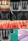
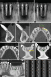
Periapical lesions with mixed radiographic appearance can have odontogenic or nonodontogenic origin. A number of neoplastic lesions either benign or malignant can present as radiolucent, radiopaque, or mixed in jaws and if present near the root apices can be misdiagnosed as odontogenic infection/etiology. The present case report describes a rare case of two elongated radiopaque structures within periapical pathology located beneath the apices of mandibular central incisors in a 26-year-old male. Further, it describes its nonsurgical and surgical endodontic management along with histological confirmation and long-term radiographic healing outcome using cone-beam computed tomography. Microscopic examination revealed the presence of dentin and cementum with fringes of periodontal ligament suggestive of tooth-like structures. No case report has yet reported tooth-like calcifications within the large periapical lesion. Biopsy of such lesions is deemed necessary to differentiate from nonodontogenic lesions which could be benign or malignant in nature.
Keywords: Apicoectomy; bacteria; calcification; cone beam computed tomography; foreign bodies; periapical granuloma.
Copyright: © 2024 Journal of Conservative Dentistry and Endodontics.
Conflict of interest statement
There are no conflicts of interest.
Figures


References
-
- Vieira CC, Pappen FG, Kirschnick LB, Cademartori MG, Nóbrega KH, do Couto AM, et al. A retrospective Brazilian multicenter study of biopsies at the periapical area: Identification of cases of nonendodontic periapical lesions. J Endod. 2020;46:490–5. - PubMed
-
- Bornstein MM, Bingisser AC, Reichart PA, Sendi P, Bosshardt DD, von Arx T. Comparison between radiographic (2-dimensional and 3-dimensional) and histologic findings of periapical lesions treated with apical surgery. J Endod. 2015;41:804–11. - PubMed
-
- Nair PN. Pathogenesis of apical periodontitis and the causes of endodontic failures. Crit Rev Oral Biol Med. 2004;15:348–81. - PubMed
-
- Sirotheau Corrêa Pontes F, Paiva Fonseca F, Souza de Jesus A, Garcia Alves AC, Marques Araújo L, Silva do Nascimento L, et al. Nonendodontic lesions misdiagnosed as apical periodontitis lesions: Series of case reports and review of literature. J Endod. 2014;40:16–27. - PubMed
Publication types
LinkOut - more resources
Full Text Sources
