. 2024 Sep 30:11:1436356.
doi: 10.3389/fvets.2024.1436356. eCollection 2024.

Computed tomographic characteristics of craniomandibular osteopathy in 20 dogs

Affiliations

Computed tomographic characteristics of craniomandibular osteopathy in 20 dogs

L A Pérez López et al. Front Vet Sci. .

Abstract

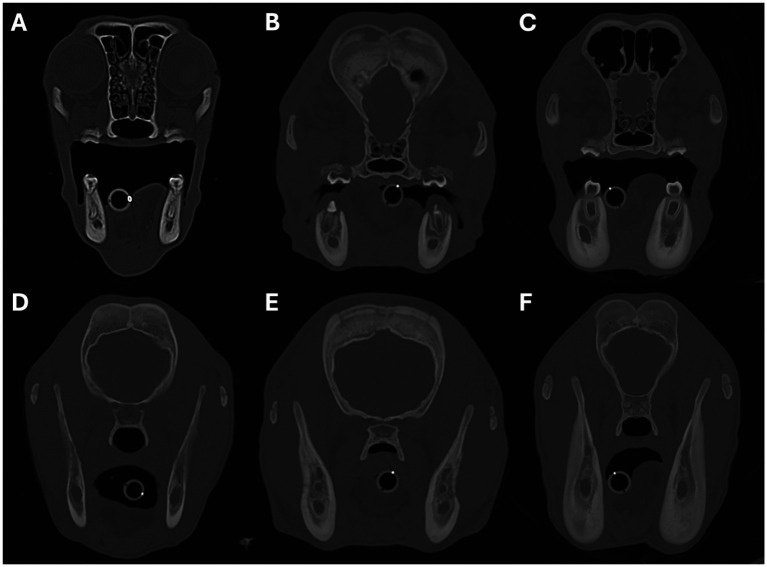
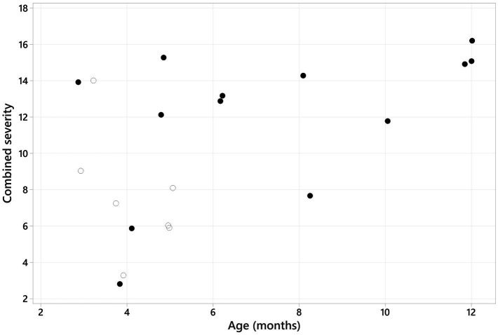
Craniomandibular osteopathy (CMO) is a proliferative, self-limiting, non-neoplastic disease of growing dogs characterised by excessive new bone formation on the skull and mandible. The radiographic findings of CMO are well described; however, limited reports of the computed tomographic (CT) appearance are available. This paper aims to characterise the spectrum of CT findings that can occur with CMO. The study is retrospective, descriptive, multicenter, and includes 20 cases. Age at presentation ranged from 6 weeks to 12 months, with no sex predisposition. Scottish terriers were overrepresented (65%); other breeds included Cairn terrier, Jack Russell terrier, Staffordshire bull terrier, labrador retriever, golden retriever, akita and Slovakian rough-haired pointer (one of each breed). Terrier breeds represented 80% (16/20) of the patient cohort. Mandibular osteoproliferation was present in all patients (marked in 80%, bilateral in 95%), affecting the rostral mandible in 25%, body in 85%, and ramus in 80%. Tympanic bulla osteoproliferation was present in 60% (12/20) of patients (all marked, bilateral in 75%). Cranial osteoproliferation (frontal, parietal, temporal, occipital bones or maxilla, or combinations of them) was present in 90% (18/20) of patients (40% marked, 27% moderate, 33% mild). Nasopharyngeal narrowing was seen in all 12 patients with tympanic bulla osteoproliferation (67% marked, 27% moderate) and caused nearly complete occlusion in two of them. External ear canal stenosis was seen in 55% (11/20) of patients (63% marked, 37% moderate, all bilateral). Temporomandibular joint (TMJ) impingement was suspected in 83% (10/12) of patients with marked tympanic bulla osteoproliferation (75% bilateral). Osteolysis with a moth-eaten pattern was seen in the mandible of 10/20 dogs, the calvarium of 5/20 dogs, and the maxilla of 1/20 dogs (5%). Lymphadenomegaly (mandibular and medial retropharyngeal) was found in 15/20 patients (70% mild, 30% moderate). The most severe CT changes were seen in Scottish terriers. CT allows for detailed characterisation of the bony changes associated with CMO, including the effects occurring secondary to osteoproliferation surrounding the tympanic bullae such as TMJ impingement, external ear canal stenosis, and nasopharyngeal narrowing. Osteoproliferation affecting the cranium and the presence of osteolysis were seen more frequently in this study than previously reported in CMO.

Keywords: craniomandibular; external ear canal stenosis; lion’s jaw; pharyngeal stenosis; tympanic bullae and calvarian osteoproliferation.


Conflict of interest statement

The authors declare that the research was conducted in the absence of any commercial or financial relationships that could be construed as a potential conflict of interest.

Figures

Figure 1
Figure 1
Sagittal (A,D), transverse (B,E), and dorsal oblique (C,F) multiplanar reconstruction images from a 5-month-old Scottish terrier showing how the severity of the nasopharyngeal narrowing was calculated. Images A–C show measurement of the maximum cross-sectional area of the nasopharynx, just caudal to the hamulus of the pterygoid bone. Images D–F show how the minimum cross-sectional area of the nasopharynx was measured.
Figure 2
Figure 2
Transverse images showing examples of periosteal new bone formation affecting the mandibles. Images A–C show examples of osteoproliferation affecting the body of the mandible, and images D–F show osteoproliferation affecting the mandibular ramus. The severity of the osteoproliferation was graded as mild in images (A,D), moderate in images (B,E), and marked in images (C,F). Image A is from a 5-month-old Scottish terrier, B,E are from a 4-month-old Jack Russell terrier, C,F are from a 5-month-old akita, and D is from a 5-month-old Slovakian rough-haired pointer.
Figure 3
Figure 3
Transverse images showing examples of osteolysis affecting the mandibles. Images A–C show examples of osteolysis affecting the body of the mandible, and image D shows osteolysis affecting the mandibular ramus. The regions of osteolysis are denoted by the white arrowheads. Images A,B are from a 4-month-old Scottish terrier, and C,D are from a 4-month-old Jack Russell terrier.
Figure 4
Figure 4
Transverse (A–D), dorsal (E), and sagittal oblique 3D volume rendered images showing examples of marked periosteal new bone formation affecting the tympanic bullae. In images A,C, the osteoproliferation has a predominantly solid and dense appearance; whereas in image B, it has an irregular lobular appearance. Images D–F show examples of the osteoproliferation surrounding the tympanic bullae extending around and engulfing the stylohyoid bones. Images A,B are from an 8-month-old Scottish terrier, C is from a 12-month-old Scottish terrier, and D–F are from a 6-month-old Scottish terrier.
Figure 5
Figure 5
Transverse (A,C,E) and dorsal oblique (B,D,F) multiplanar reconstruction images showing examples of nasopharyngeal narrowing secondary to osteoproliferation surrounding the tympanic bullae. Images A,B are from a patient with no evidence of nasopharyngeal narrowing. Images C, D are from a patient with moderate nasopharyngeal narrowing, and images E,F are from a patient with marked nasopharyngeal narrowing. Images A, B are from a 4-month-old Scottish terrier, C,D are from a 6-month-old Scottish Terrier, and E,F are from an 8-month-old Scottish terrier.
Figure 6
Figure 6
Transverse images showing examples of external ear canal stenosis secondary to osteoproliferation surrounding the tympanic bullae. Image A shows a normal horizontal ear canal with no evidence of stenosis. The remaining images show examples of patients with mild (image B), moderate (image C), and marked (imaged D) stenosis of the horizontal ear canal. Image A is from a 3-month-old labrador retriever, B is from a 5-month-old Scottish terrier, C is from an 8-month-old Scottish terrier, and D is from a 12-month-old Scottish terrier.
Figure 7
Figure 7
Sagittal oblique (A,C) and dorsal oblique (D,F) multiplanar reconstruction images showing examples of temporomandibular joint impingement secondary to osteoproliferation surrounding the tympanic bullae. Images A,C show how the proliferative new bone formation arising from the tympanic bulla extends rostrally and closely conforms to the caudal margin of the mandibular ramus, preventing the normal hinge-like opening of the temporomandibular joint. Images D,F show how the osteoproliferation also extends along the medial aspect of the mandibular ramus. Images B,E are photographs of a macerated skull showing severe osteoproliferation affecting the tympanic bullae and mandibular rami. Images A,B,D,E are from a 12-month-old Scottish terrier, C,F are from an 8-month-old Scottish terrier.
Figure 8
Figure 8
Transverse images showing examples of periosteal new bone formation affecting the cranium. Images A–C show examples of osteoproliferation affecting the frontal bones, and images D–F show osteoproliferation affecting the parietal bones. The severity of the osteoproliferation was graded as mild in images A,D, moderate in images B,E, and marked in images C,F. Image A is from a 4-month-old Slovakian rough-haired pointer, B is from a 4-month-old Cairn terrier, C,F are from a 4-month-old Staffordshire bull terrier, D is from a 4-month-old Jack Russell terrier, and E is from an 8-month-old Scottish terrier.
Figure 9
Figure 9
Transverse images showing examples of osteolysis affecting the cranium. Images A,B show examples of osteolysis affecting the frontal bones, and images C,D show osteolysis affecting the parietal bones. Image A is from a 5-month-old golden retriever, B,D are from a 4-month-old Staffordshire bull terrier, and C is from a 4-month-old Cairn terrier.
Figure 10
Figure 10
Scatter plot graph showing the combined severity (sum of the scores for each CT feature) plotted against the age of the patient. A positive Spearman rank correlation was identified (rs = 0.481, p = 0.032). The solid black dots represent Scottish terriers, and the empty/white dots represent the remaining breeds.
Figure 11
Figure 11
Boxplot showing the combined severity in Scottish terriers (blue box) versus the other breeds (red box). There was a significantly higher combined severity in Scottish terriers compared to other breeds (p = 0.038).

References

    1. Vagt J, Distl O. Complex segregation analysis of craniomandibular osteopathy in Deutsch Drahthaar dogs. Vet J. (2018) 231:30–2. doi: 10.1016/j.tvjl.2017.11.008 - DOI - PubMed
    1. Alexander JW. Selected skeletal dysplasias: craniomandibular osteopathy, multiple cartilaginous exostoses, and hypertrophic osteodystrophy. Vet Clin North Am Small Anim Pract. (1983) 13:55–70. doi: 10.1016/S0195-5616(83)50004-1, PMID: - DOI - PubMed
    1. Pettitt R, Fox R, Comerford EJ, Newitt A. Bilateral angular carpal deformity in a dog with craniomandibular osteopathy. Vet Comp Orthop Traumatol. (2012) 25:149–54. doi: 10.3415/VCOT-11-02-0022 - DOI - PubMed
    1. Padgett GA, Mostosky UV. Animal model: the mode of inheritance of craniomandibular osteopathy in West Highland white terrier dogs. Am J Med Genet. (1986) 25:9–13. doi: 10.1002/ajmg.1320250103, PMID: - DOI - PubMed
    1. Riser WH, Parkes LJ, Shirer JF. Canine Craniomandibular osteopathy. J American Vet Radiol Society. (1967) 8:23–31. doi: 10.1111/j.1740-8261.1967.tb01069.x - DOI

LinkOut - more resources