Observational Study
. 2024 Nov 5;13(21):e036065.
doi: 10.1161/JAHA.124.036065. Epub 2024 Oct 25.

Stent Underexpansion Is an Underestimated Cause of Intrastent Restenosis: Insights From RESTO Registry

Affiliations
Observational Study

Stent Underexpansion Is an Underestimated Cause of Intrastent Restenosis: Insights From RESTO Registry

Géraud Souteyrand et al. J Am Heart Assoc. .

Abstract

Background: Despite improvement in devices, in-stent restenosis remains a frequent and challenging complication of percutaneous coronary interventions.

Methods and results: The RESTO (Morphological Parameters of In-Stent Restenosis Assessed and Identified by OCT [Optical Coherence Tomography]; study NCT04268875) was a prospective multicenter registry including patients presenting with coronary syndromes related to in-stent restenosis. All patients underwent preintervention OCT analysis, which led to analysis of in-stent restenosis phenotype, number of strut layers, and presence of stent underexpansion. The primary end point was the in-stent restenosis type according to the OCT morphological classification. The 1-year incidence of target vessel failure (a composite of death from cardiac causes, target-vessel myocardial infarction, or ischemia-driven target-vessel revascularization) was assessed. The study included 297 patients. The culprit stent was a drug-eluting stent in 74.2% of cases. OCT analysis revealed the presence of neoatherosclerosis in 57% (52% calcified), neointimal hyperplasia in 43% (58% homogeneous), stent underexpansion (minimal stent area <4.5 mm2) in 43%, and multiple stent layers in 30%. The prepercutaneous coronary intervention OCT analysis modified the operator's strategy for management in 30% of cases. Treatment involved drug-eluting stent implantation in 61.6% and drug-eluting balloon angioplasty in 36.1% of cases with only 63.2% optimal results. The 1-year target vessel failure incidence was 11% (95% CI, 9%-13%). Residual postpercutaneous coronary intervention stent underexpansion was associated with significantly higher target vessel failure incidence (19% [95% CI, 14%-24%] versus 7% [95% CI, 5-9], P=0.01).

Conclusions: OCT identified neoatherosclerosis and neointimal hyperplasia in comparable proportions. Stent underexpansion was frequent and favored subsequent adverse clinical outcomes.

Keywords: in‐stent restenosis; optical coherence tomography; underexpansion.


Figures

Figure 1
Figure 1. Representative examples of different ISR patterns analyzed by OCT.
A, Homogeneous NIH; B, heterogeneous NIH; C, NIH related to multiple stent layers and device underexpansion; D, noncalcified fibrolipidic neoatherosclerosis; E, calcified neoatherosclerosis; F, intrastent calcified nodule; G, an example of severe stent underexpansion; H, focal and diffuse lesion pattern by longitudinal OCT analysis. ISR indicates in‐stent retinosis; NIH, neointimal hyperplasia; OCT, optical coherence tomography; SA, stent area; and SD, stent diameter.
Figure 2
Figure 2. Prevalence of different ISR patterns in the RESTO cohort (N=297 patients).
Calcified neoatherosclerosis: n=85 (29%); noncalcified neoatherosclerosis: n=83 (28%); homogeneous NIH: n=74 (25%); layered NIH: n=55 (18%). ISR indicates in‐stent retinosis; NIH, neointimal hyperplasia; and RESTO, Morphological Parameters of In‐Stent Restenosis Assessed and Identified by OCT [Optical Coherence Tomography].
Figure 3
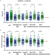
Figure 3. Variations of stent expansion according to crude MSA analysis (A) or geometrical analysis (B) before and after culprit lesion treatment in the whole cohort and among the different ISR patterns.
Whisker box represent median and interquartile range. Homogeneous NIH (n=74); Layered NIH (n=55); Ca2+ NA (n=85); non‐Ca2+ NA neoatherosclerosis (n=83)/*P<0.01; # P<0.05 by paired Student t test. Ca2+ indicates calcified; ISR, in‐stent retinosis; MSA, minimal stent area; NIH, neointimal hyperplasia; and PCI, percutaneous coronary intervention.
Figure 4
Figure 4. Therapeutic change after OCT.
CABG indicates coronary artery bypass graft; DEB, drug eluting balloon; DES, drug eluting stent; OCT, optical coherence tomography; and PCI, percutaneous coronary intervention.
Figure 5
Figure 5. Relation between OCT geometrical expansion and MSA post PCI.
MSA indicates minimal stent area; OCT, optical coherence tomography; and POBA, plain old balloon angiography.
Figure 6
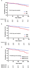
Figure 6. Kaplan–Meier estimates of the survival free from TVF through the first year among different groups of patients: patients with neoatherosclerosis or NIH (A), patients treated with DEB or DES (B), patients with or without MSA <4.5 mm2 post ISR treatment according to crude MSA (C).
DEB indicates drug eluting balloon; DES, drug eluting stent; ISR, in‐stent retinosis; MSA, minimal stent area; NIH, neointimal hyperplasia; and TVF, target vessel failure.
Figure 7
Figure 7. The proposed RESTO algorithm for OCT‐guided ISR management.
DCB indicates drug coated balloon; DES, drug eluting stent; ISR, intrastent restenosis; IVL, intravascular lithotripsy; NCB, noncompliant balloon; OCT, optical coherence tomography; OPN, ultra high pressure NCB; PCI, percutaneous coronary intervention; RESTO, Morphological Parameters of In‐Stent Restenosis Assessed and Identified by OCT [Optical Coherence Tomography]; and Rot Ath, rotational atherectomy.

References

    1. Neumann F‐J, Sousa‐Uva M, Ahlsson A, Alfonso F, Banning AP, Benedetto U, Byrne RA, Collet J‐P, Falk V, Head SJ, et al. 2018 ESC/EACTS Guidelines on myocardial revascularization. Eur Heart J. 2019;40:87–165. doi: 10.1093/eurheartj/ehy394 - DOI - PubMed
    1. Lee MS, Banka G. In‐stent restenosis. Interv Cardiol Clin. 2016;5:211–220. doi: 10.1016/j.iccl.2015.12.006 - DOI - PubMed
    1. Bønaa KH, Mannsverk J, Wiseth R, Aaberge L, Myreng Y, Nygård O, Nilsen DW, Kløw N‐E, Uchto M, Trovik T, et al. Drug‐eluting or bare‐metal stents for coronary artery disease. N Engl J Med. 2016;375:1242–1252. doi: 10.1056/NEJMoa1607991 - DOI - PubMed
    1. Yıldız A, Yıldız C, Oduncu V, Çimen AO. A comparison of drug‐eluting stent versus balloon angioplasty in patients with bare‐metal stent instent restenosis: 5 year outcomes. Int J Cardiovasc Acad. 2016;2:1–5. doi: 10.1016/j.ijcac.2016.02.003 - DOI
    1. Xiu W‐J, Yang H‐T, Zheng Y‐Y, Ma Y‐T, Xie X. Drug‐eluting balloons versus second‐generation drug‐eluting stents for treating in‐stent restenosis in coronary heart disease after PCI: a meta‐analysis. Cardiol Res Pract. 2018;2018:1–14. doi: 10.1155/2018/7658145 - DOI - PMC - PubMed

Publication types

MeSH terms

LinkOut - more resources