. 2025 Jan 1;63(1):118-131.
doi: 10.1097/SHK.0000000000002476. Epub 2024 Oct 18.

HOUSING TEMPERATURE ALTERS BURN-INDUCED HYPERMETABOLISM IN MICE

Affiliations

HOUSING TEMPERATURE ALTERS BURN-INDUCED HYPERMETABOLISM IN MICE

Meagan Scott Kingren et al. Shock. .

Abstract

Mice used in biomedical research are typically housed at ambient temperatures (22°C-24°C) below thermoneutrality (26°C-31°C). This chronic cold stress triggers a hypermetabolic response that may limit the utility of mice in modeling hypermetabolism in response to burns. To evaluate the effect of housing temperature on burn-induced hypermetabolism, mice were randomly assigned to receive sham, small, or large scald burns. Mice recovered for 21 days in metabolic phenotyping cages at 24°C or 30°C. Regardless of sex or sham/burn treatment, mice housed at 24°C had greater total energy expenditure ( P < 0.001), which was largely attributable to greater basal energy expenditure when compared to mice housed at 30°C ( P < 0.001). Thermoneutral housing (30°C) altered adipose tissue mass in a sex-dependent manner. Compared to sham and small burn groups, large burns resulted in greater water vapor loss, regardless of housing temperature ( P < 0.01). Compared to sham, large burns resulted in greater basal energy expenditure and total energy expenditure in mice housed at 24°C; however, this hypermetabolic response to large burns was blunted in female mice housed at 30°C, and absent in male mice housed at 30°C. Locomotion was significantly reduced in mice with large burns compared to sham and small burn groups, irrespective of sex or housing temperature ( P < 0.05). Housing at 30°C revealed sexual dimorphism in terms of the impact of burns on body mass and composition, where males with large burns displayed marked cachexia, whereas females did not. Collectively, this study demonstrates a sex-dependent role for housing temperature in influencing energetics and body composition in a rodent model of burn trauma.


Conflict of interest statement

The authors report no conflicts of interest.

Figures

Figure 1:
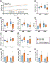
Figure 1:. Housing temperature alters whole body energy expenditure and animal behavior.
Mice were housed at either 24°C or 30°C for 21 days during which energy expenditure was constantly monitored. Energy expenditure for each hour of the experiment (A), average daily TEE (B), and average daily BEE (C) were significantly reduced in animals housed at 30°C versus 24°C. BEE and TEE were then graphed together (D-E). Each point represents a different animal. Daily movement of each animal about its cage without the presence of a running wheel was reported (F). There were no significant differences in average daily locomotion as a result of housing temperature, though 24°C housed females ambulated more than their male counterparts (G). Daily food intake was reported (H). Overall, animals housed at 24°C had greater food intake than 30°C housed animals (H). Fecal output, regardless of sex, was greater in animals housed at 24°C (I). The energy of the feces (GEF) was less in 30°C housed animals, while male animals at both temperatures had increased GEF (J). Each point represents a different animal. TEE, total energy expenditure; AEE, active energy expenditure; BEE, basal energy expenditure; GEF, gross energy of feces; significant effect of sex is represented by #; significant effect of temperature is represented by †; one symbol, (P < 0.05); two symbols, (P < 0.01); three symbols, (P < 0.001).
Figure 2:
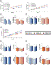
Figure 2:. Impact of housing temperature on body composition in sham mice.
Body mass was monitored throughout the experiment (A). Prior to sham injury and euthanasia, each animal’s body composition was evaluated using DEXA. Change over the course of the 21-day experiment is shown for body mass (B), lean mass (C), fat mass (D) bone mineral density (E), relative lean mass (F), and relative fat mass (G) are shown. Following euthanasia, fat pads were isolated, and iWAT (H), gWAT (I), and BAT (J) masses are reported. Each point represents a different animal. BAT, brown adipose tissue; iWAT, white adipose tissue; gWAT, gonadal white adipose tissue; fat pads were normalized to body mass; significant effect of sex is represented by #; significant effect of temperature is represented by †; one symbol, (P < 0.05); two symbols, (P < 0.01); three symbols, (P < 0.001).
Figure 3.
Figure 3.. Burn injury triggers a moderate hypermetabolic response in mice.
Hourly energy expenditure was reported (A), and average daily TEE analysis revealed temperature was the major determinant of metabolism (B). Parameters were then graphed separately for cold and thermoneutral housing temperature conditions. Daily TEE (C) and BEE (D) are shown for each of the 21 days. Average daily BEE (E) and average daily relative BEE (F) are shown. Each point represents a different animal. TEE, total energy expenditure; BEE, basal energy expenditure; Significant effect of burn injury is represented by *; significant effect of temperature is represented by †; one symbol, (P < 0.05); two symbols, (P < 0.01); three symbols, (P < 0.001).
Figure 4.
Figure 4.. Burn injury triggers evaporative heat loss.
Evaporative heat loss was measured each day after burn injury (A). Large burns caused increased evaporative heat loss in both 24°C and 30°C housed mice (B). Average daily evaporative heat loss was not altered by housing temperature but by burn severity (C). Each point represents a different animal. Significant effect of burn injury *; significant effect of sex is represented by #; significant effect of temperature is represented by †; one symbol, (P < 0.05); two symbols, (P < 0.01); three symbols, (P < 0.001).
Figure 5:
Figure 5:. Burn injury influences locomotion and feeding characteristics.
Ambulatory movement was reported daily (A). There were no significant differences in average daily locomotion as a result of housing temperature, though animals subjected to large burns moved significantly less than their small burn and sham counterparts (B). Burn injury also reduced ambulatory speed (C). Daily food intake was reported (D). Overall, animals housed at 24°C had greater food intake and GEI than 30°C housed animals, but burn injury had minimal effects (E). Fecal output (F) increased in 24°C housed female housed mice subjected to large burn injury. At 30°C, female mice did not undergo any changes as a result of burn injury, though all female mice consumed less food than their male counterparts. The energy of the feces was calculated (G). GEF followed the same trends as fecal output (H). Each point represents a different animal. GEI, gross energy intake; GEF, gross energy of feces; significant effect of burn injury is represented by *; significant effect of sex is represented by #; significant effect of temperature is represented by †; one symbol, (P < 0.05); two symbols, (P < 0.01); three symbols, (P < 0.001).
Figure 6:
Figure 6:. Burn injury, temperature, and sex all influence body composition.
Daily body mass is reported for male and female mice at both 24°C and 30°C (A). Each animal’s body composition was evaluated using DEXA prior to burn injury and again on the morning of euthanasia for tissue collection. The percent changes over the 21-day experiment for body mass (B), absolute lean mass (C), absolute fat mass (D), relative lean mass (E), and relative fat mass (F) are shown. Following euthanasia, fat pads were isolated, and iWAT (G), gWAT (H), and BAT (I) masses are reported as values normalized to body mass. The change in BMD (J) and BMC (K) were also calculated using DEXA scans before injury and euthanasia. Each point represents a different animal. BAT, brown adipose tissue; iWAT, white adipose tissue; gWAT, gonadal white adipose tissue; BMD, bone mineral density; BMC, bone mineral content; significant effect of burn injury is represented by *; significant effect of sex is represented by #; one symbol, (P < 0.05); two symbols, (P < 0.01); three symbols, (P < 0.001).
Figure 7.
Figure 7.. Large burn triggers moderate hypermetabolism.
Secondary analyses were completed for TEE (A), BEE (B), and VH2O loss (C) to evaluate the effect of large burn injury. Main effects were evaluating using unpaired t-tests. Significant effect of burn injury is represented by *; one symbol, (P < 0.05); two symbols, (P < 0.01); three symbols, (P < 0.001).

References

    1. National Hospital Ambulatory Medical Care Survey: 2021 Emergency Department Summary Tables. Published online 2021.
    1. Caldwell FT, Bowser BH, Crabtree JH. The effect of occlusive dressings on the energy metabolism of severely burned children. Ann Surg. 1981;193(5):579–591. doi: 10.1097/00000658-198105000-00007 - DOI - PMC - PubMed
    1. Wilmore DW, Mason AD, Johnson DW, Pruitt BA. Effect of ambient temperature on heat production and heat loss in burn patients. J Appl Physiol. 1975;38(4):593–597. doi: 10.1152/jappl.1975.38.4.593 - DOI - PubMed
    1. Porter C, Tompkins RG, Finnerty CC, Sidossis LS, Suman OE, Herndon DN. The Metabolic Stress Response to Burn Trauma: Current Understanding and Therapies. Lancet Lond Engl. 2016;388(10052):1417–1426. doi: 10.1016/S0140-6736(16)31469-6 - DOI - PMC - PubMed
    1. Jeschke MG, Gauglitz GG, Kulp GA, et al. Long-term persistance of the pathophysiologic response to severe burn injury. PloS One. 2011;6(7):e21245. doi: 10.1371/journal.pone.0021245 - DOI - PMC - PubMed