. 2024 Dec 1;213(11):1644-1655.
doi: 10.4049/jimmunol.2300858.

Single-cell Sequencing of Circulating Human Plasmablasts during Staphylococcus aureus Bacteremia

Affiliations

Single-cell Sequencing of Circulating Human Plasmablasts during Staphylococcus aureus Bacteremia

Priscilla F Kerkman et al. J Immunol. .

Abstract

Staphylococcus aureus is the major cause of healthcare-associated infections, including life-threatening conditions as bacteremia, endocarditis, and implant-associated infections. Despite adequate antibiotic treatment, the mortality of S. aureus bacteremia remains high. This calls for different strategies to treat this infection. In past years, sequencing of Ab repertoires from individuals previously exposed to a pathogen emerged as a successful method to discover novel therapeutic monoclonal Abs and understand circulating B cell diversity during infection. In this paper, we collected peripheral blood from 17 S. aureus bacteremia patients to study circulating plasmablast responses. Using single-cell transcriptome gene expression combined with sequencing of variable heavy and light Ig genes, we retrieved sequences from >400 plasmablasts revealing a high diversity with >300 unique variable heavy and light sequences. More than 200 variable sequences were synthesized to produce recombinant IgGs that were analyzed for binding to S. aureus whole bacterial cells. This revealed four novel monoclonal Abs that could specifically bind to the surface of S. aureus in the absence of Ig-binding surface SpA. Interestingly, three of four mAbs showed cross-reactivity with Staphylococcus epidermidis. Target identification revealed that the S. aureus-specific mAb BC153 targets wall teichoic acid, whereas cross-reactive mAbs BC019, BC020, and BC021 target lipoteichoic acid. All mAbs could induce Fc-dependent phagocytosis of staphylococci by human neutrophils. Altogether, we characterize the active B cell responses to S. aureus in infected patients and identify four functional mAbs against the S. aureus surface, of which three cross-react with S. epidermidis.


Figures

Figure 1
Figure 1. SAB patients have plasmablasts that produce S aureus recognizing antibodies.
A) The frequency of CD19+ B cells in total live single PBMC (left) and CD3-CD14-CD19+/dimCD27+CD38+CD20- plasmablasts in CD3-CD14-CD19+/dim total B cells (right) of patients ((day 8-21, n=12) or (>day 30, n=5)) and healthy donors (HD, n=6). The SAB patients were divided into two groups, day 8-21 or >day 30, based on the day of the blood draw that followed the first S. aureus positive blood culture. Each dot represents one donor and the colored dots refer to patients 1-3 and 6, which were the focus of further analysis. (B) Quantification of total IgG levels in culture supernatants. 50.000 PBMCs from patients or healthy donors (HD) were cultured for 7 days without additional stimuli. ELISA was used to assess total IgG levels in the resulting supernatants. The grey area shows interpolated values below standard curve. Every dot represents one well. (C, D) Quantification of S. aureus-specific IgG in PBMC culture supernatants. Binding of mAbs to S. aureus Newman ΔSpA/ΔSbi was detected using anti human IgG AlexaFluor647 and analyzed using flow cytometry. Bacteria were gated using forward and side scatter (gating strategy in C, black oval) to determine the geomean value for the bacterial population. For each well, the geomean value of the gated bacterial population is divided by the geomean of an empty control well (D). The dotted line represents control value. The colored line represents median, n=17-20 wells per donor.
Figure 2
Figure 2. Sequence analysis of patient plasmablasts.
A) Left: representative plot of t-SNE clustering of expression profiles. The different colors are used to separate the six clusters with similar mRNA expression profiles of donor 1. Upper right panel, cells with an expression pattern CD27+CD38+CD20- in pink, and lower right panel shows the log2 CD27 expression level of cells. B) Isotype distribution of all CD27+CD38+CD20- unique plasmablast (PB) clonotypes derived from six donors. C) The CDR3 length of heavy chain C and number of somatic hypermutations (SHM) in the D27+CD38+CD20- plasmablast clonotypes. Clonotypes grouped per patient (in color) that could be assigned to a patient using the oligonucleotide tag (expression >7log2, for the hashed sort) and the clonotypes that could not be allocated in black (unknown). For patient 1 & patient 6 clonotypes from enriched and hashed sort were combined into one column. Every dot is one clonotype.
Figure 3
Figure 3. Identification of 4 unique antibodies recognizing S. aureus.
A) Left: Representative histogram of a typical binding experiment in which antibody supernatants were incubated with S. aureus and then detected with αIgG by flow cytometry. Light grey filled: buffer control; dark grey line: non-binding supernatant; black line: binding antibody in supernatant. Right: Relative binding of antibodies in 224 supernatants to clinical isolates from six patients. Binding was defined as relative binding >2 (above dotted lines). Each dot is one supernatant/mAb. B) Supernatants that contain IgGs against one of the clinical isolates were tested for relative binding to S. aureus Newman WT versus Δspa/sbi. In green: supernatants binding to Newman Δspa/sbi; lower right quadrant supernatants binding only to Newman WT (Spa/Sbi binding to supernatants); lower left quadrant not binding to Newman WT (but binding to at least one of the clinical isolates). C) Alignment of CDR3 HC and LC amino acid sequences of BC019, BC020, BC021 and BC153. D) Relative binding of four mAbs (1 µg/ml) to clinical isolates from six patients, Newman WT, and Δspa/sbi (New KO). A-B,D) For all strains, buffer controls were included and used to calculate relative binding of each supernatant/mAb by dividing the geomean of a supernatant/mAb by the geomean of the buffer control binding the same isolate. E) Binding of 1 µg/ml mAbs to SpA-BKK (a variant that lacks IgG-Fc binding) using ELISA. SpA-BKK binds to BC019 and BC021 but not BC020. (n=1).
Figure 4
Figure 4. Binding of newly identified mAbs to S. aureus and S epidermidis.
Newly identified antibodies were compared to anti-WTA (4497) (S. aureus specific), anti-SDR (rF1) (known to bind both S. aureus and S. epidermidis (33) and anti-DNP antibody as a control. Binding titration curve of mAbs (A,C) or binding of mAbs at 1 μg/ml (B). Binding to S. aureus Newman Δspa/sbi (A), 8 S. epidermidis clinical isolates (B) and S. epidermidis clinical isolate (N4843) (C). Binding of mAbs was detected using anti-human IgG AlexaFluor647 and analyzed using flow cytometry. Bacteria were gated using forward and side scatter to determine the geomean value for the bacterial population. The average of three independent experiments is plotted; (A,C) includes the ± SD; (B) only the average of three independent experiments.
Figure 5
Figure 5. Target identification of newly identified mAbs.
Binding of antibodies to purified staphylococcal surface carbohydrates. (A-C) ELISA plates were coated with purified PG (A, 2 µg/mL), PNAG (B, 0.6 µg/mL) or LTA (C, 2 µg/mL) and incubated with 1 µg/mL IgG3 of the four newly identified antibodies. Antibody binding was detected using anti-human-IgG-HRP antibodies. Anti-PG (M130), anti-PNAG (F598) and anti-LTA (A120) (all as IgG3 at 1 µg/ml) were used as positive control. Anti β-GlcNAc-WTA (4497) and anti-DNP and served as negative controls. (D) Binding of 1 µg/mL IgG3 antibodies to synthetic WTA beads, either only ribitol phosphate backbone (RboP), or modified with TarM or TarS to obtain α- or β-GlcNAc modifications respectively. Antibody binding was detected with goat-anti-human-kappa-AF488 and measured by flow cytometry. Antibodies targeting α-GlcNac-WTA (4461) and β-GlcNAc-WTA (4497) were included as positive controls. (A-C) Data represent mean ± SD of two independent experiments. (B) Data represent mean ± SD of three independent experiments.
Figure 6
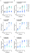
Figure 6. Phagocytosis of S. aureus and S. epidermidis by the identified antibodies.
Newly identified antibodies were compared to anti-WTA (4497) (A-D), anti-SDR and control anti-DNP antibody. Phagocytosis of S. aureus Newman Δspa/sbi (A-B) S. aureus N0280 (C-D) and S. epidermidis (E-F) by human neutrophils MOI 10:1 in absence (A,C,E) or presence (B,D,F) of complement. mAmetrine expressing S. aureus Newman Δspa/sbi, or FITC-labelled S. aureus N0280 or S. epidermidis (N4843) were pre-incubated with a concentration range of mAbs in RPMI-H only or including 1% IgG/IgM-depleted pooled normal human serum. After 15 min incubation neutrophils were added for an additional 15 min at 37°C. Phagocytosis of fluorescent bacteria was analyzed by flow cytometry and is expressed as the mAmetrine/FITC G=geomean of gated neutrophils. Plotted is the average of three independent experiments ± SD (A-B, E-F) or n=1 (C-D).

References

    1. Lister JL, Horswill AR. Staphylococcus aureus biofilms: recent developments in biofilm dispersal. Front Cell Infect Microbiol. 2014;4:178. doi: 10.3389/fcimb.2014.00178. - DOI - PMC - PubMed
    1. Lowy FD. Staphylococcus aureus infections. N Engl J Med. 1998;339:520–32. - PubMed
    1. Otto M. Staphylococcus epidermidis--the “accidental” pathogen. Nat Rev Microbiol. 2009;7:555–67. doi: 10.1038/nrmicro2182. - DOI - PMC - PubMed
    1. Motley MP, Banerjee K, Fries BC. Monoclonal antibody-based therapies for bacterial infections. Curr Opin Infect Dis. 2019;32:210–216. doi: 10.1097/QCO.0000000000000539. - DOI - PMC - PubMed
    1. Seixas AMM, Sousa SA, Leitão JH. Antibody-Based Immunotherapies as a Tool for Tackling Multidrug-Resistant Bacterial Infections. Vaccines (Basel) 2022;10 doi: 10.3390/vaccines10111789. - DOI - PMC - PubMed

LinkOut - more resources