Review
. 2024 Oct 10;13(20):1675.
doi: 10.3390/cells13201675.

Autophagy-Mediated Cellular Remodeling during Terminal Differentiation of Keratinocytes in the Epidermis and Skin Appendages

Affiliations
Review

Autophagy-Mediated Cellular Remodeling during Terminal Differentiation of Keratinocytes in the Epidermis and Skin Appendages

Leopold Eckhart et al. Cells. .

Abstract

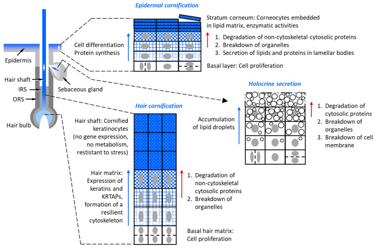
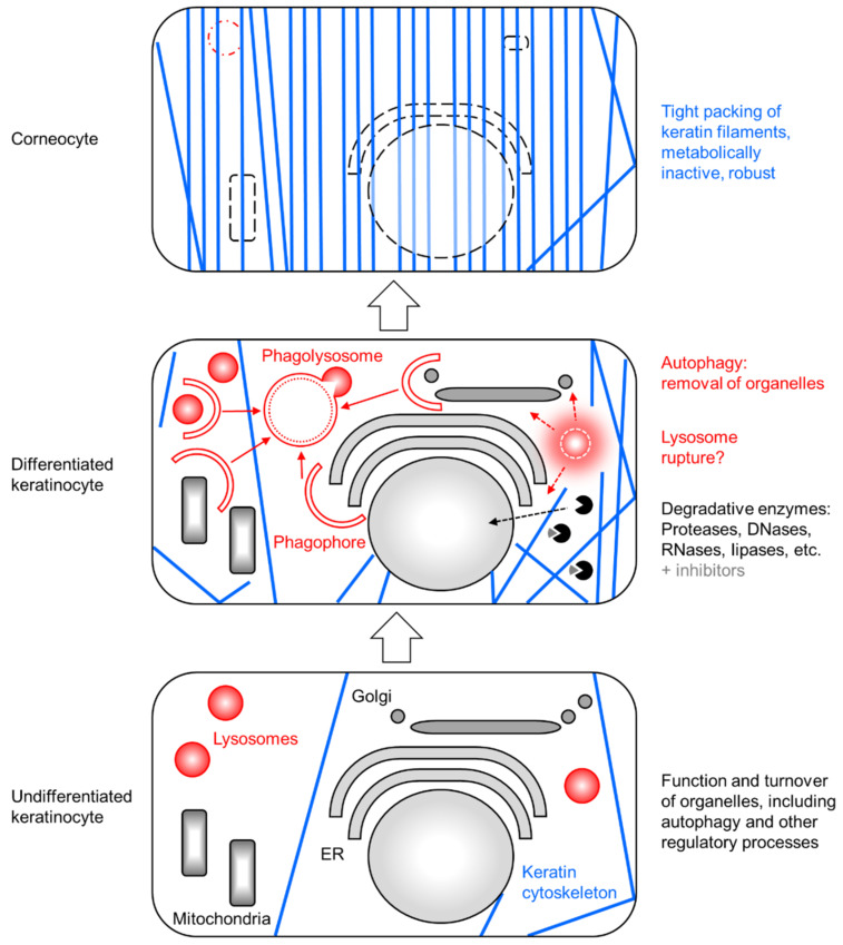
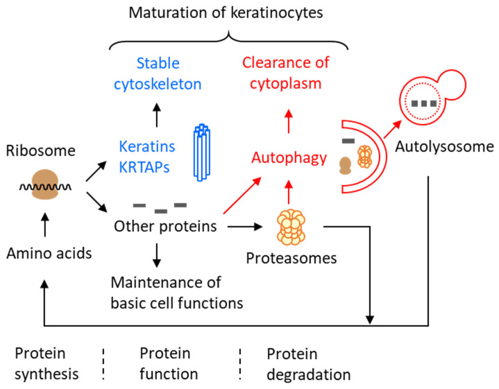
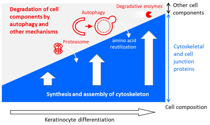
The epidermis of the skin and skin appendages, such as nails, hair and sebaceous glands, depend on a balance of cell proliferation and terminal differentiation in order to fulfill their functions at the interface of the body and the environment. The differentiation of epithelial cells of the skin, commonly referred to as keratinocytes, involves major remodeling processes that generate metabolically inactive cell remnants serving as building blocks of the epidermal stratum corneum, nail plates and hair shafts. Only sebaceous gland differentiation results in cell disintegration and holocrine secretion. A series of studies performed in the past decade have revealed that the lysosome-dependent intracellular degradation mechanism of autophagy is active during keratinocyte differentiation, and the blockade of autophagy significantly alters the properties of the differentiation products. Here, we present a model for the autophagy-mediated degradation of organelles and cytosolic proteins as an important contributor to cellular remodeling in keratinocyte differentiation. The roles of autophagy are discussed in comparison to alternative intracellular degradation mechanisms and in the context of programmed cell death as an integral end point of epithelial differentiation.

Keywords: apoptosis; autophagy; cornification; hair; keratin; keratinocytes; nail; protease; sebaceous; skin barrier.


Conflict of interest statement

The authors declare no conflicts of interest.

Figures

Figure 1
Figure 1
Degradation processes contribute to the terminal differentiation of keratinocytes in the epidermis, hair shafts and sebaceous glands. The increasing concentration of cytoskeletal proteins is highlighted by blue texture of cells (indicated by rectangles). The nucleus is depicted as a gray oval. In the schematic depiction of sebaceous gland cells, white circles indicate lipid droplets. IRS, inner root sheath; ORS, outer root sheath. This figure is adapted from a paper recently published by us under a Creative Commons CC-BY 4.0 license (open access) license [87].
Figure 2
Figure 2
Cell remodeling by autophagy in keratinocytes. Three cells representing differentiation stages of keratinocytes are schematically depicted. The degradation of organelles in differentiated keratinocytes involves an interplay between autophagy and degradative enzymes that are either released from lysosomes upon rupture of these organelles or localize to other organelles or the cytosol, where some of them are bound by proteinaceous inhibitors. ER, endoplasmic reticulum.
Figure 3
Figure 3
Protein degradation by autophagy in differentiating keratinocytes. This schematic is based on results of studies of nail plates and hair shafts from wild-type and epithelial Atg7-deficient mice. KRTAPs, keratin-associated proteins. This figure is adapted from a paper recently published by us under a Creative Commons CC-BY 4.0 license (open access) license [87].
Figure 4
Figure 4
The synthesis of the cytoskeleton is accompanied by the degradation of other cell components during the terminal differentiation of keratinocytes.

Similar articles

Cited by

References

    1. Hsu Y.C., Fuchs E. Building and maintaining the skin. Cold Spring Harb. Perspect. Biol. 2022;14:a040840. doi: 10.1101/cshperspect.a040840. - DOI - PMC - PubMed
    1. Watt F.M. Terminal differentiation of epidermal keratinocytes. Curr. Opin. Cell Biol. 1989;1:1107–1115. doi: 10.1016/S0955-0674(89)80058-4. - DOI - PubMed
    1. Fuchs E. Epidermal differentiation: The bare essentials. J. Cell Biol. 1990;111:2807–2814. doi: 10.1083/jcb.111.6.2807. - DOI - PMC - PubMed
    1. Wertz P. Epidermal lamellar granules. Skin Pharmacol. Physiol. 2018;31:262–268. doi: 10.1159/000491757. - DOI - PubMed
    1. den Hollander L., Han H., de Winter M., Svensson L., Masich S., Daneholt B., Norlén L. Skin lamellar bodies are not discrete vesicles but part of a tubuloreticular network. Acta Derm. Venereol. 2016;96:303–308. doi: 10.2340/00015555-2249. - DOI - PubMed

Publication types

LinkOut - more resources