. 2024 Oct 15;13(20):1703.
doi: 10.3390/cells13201703.

Accelerated Aging Effects Observed In Vitro after an Exposure to Gamma-Rays Delivered at Very Low and Continuous Dose-Rate Equivalent to 1-5 Weeks in International Space Station

Affiliations

Accelerated Aging Effects Observed In Vitro after an Exposure to Gamma-Rays Delivered at Very Low and Continuous Dose-Rate Equivalent to 1-5 Weeks in International Space Station

Juliette Restier-Verlet et al. Cells. .

Abstract

Radiation impacting astronauts in their spacecraft come from a "bath" of high-energy rays (0.1-0.5 mGy per mission day) that reaches deep tissues like the heart and bones and a "stochastic rain" of low-energy particles from the shielding and impacting surface tissues like skin and lenses. However, these two components cannot be reproduced on Earth together. The MarsSimulator facility (Toulouse University, France) emits, thanks to a bag containing thorium salts, a continuous exposure of 120 mSv/y, corresponding to that prevailing in the International Space Station (ISS). By using immunofluorescence, we assessed DNA double-strand breaks (DSB) induced by 1-5 weeks exposure in ISS of human tissues evoked above, identified at risk for space exploration. All the tissues tested elicited DSBs that accumulated proportionally to the dose at a tissue-dependent rate (about 40 DSB/Gy for skin, 3 times more for lens). For the lens, bones, and radiosensitive skin cells tested, perinuclear localization of phosphorylated forms of ataxia telangiectasia mutated protein (pATM) was observed during the 1st to 3rd week of exposure. Since pATM crowns were shown to reflect accelerated aging, these findings suggest that a low dose rate of 120 mSv/y may accelerate the senescence process of the tested tissues. A mathematical model of pATM crown formation and disappearance has been proposed. Further investigations are needed to document these results in order to better evaluate the risks related to space exploration.

Keywords: ATM protein; DNA double-strand breaks; accelerated aging; astronauts; risks; space radiation.


Conflict of interest statement

The authors declare no conflicts of interest.

Figures

Figure 1
Figure 1
Number of γH2AX foci per cell was assessed by immunofluorescence in the indicated cell lines and at the number of weeks of exposure at 120 mSv/y. The panel (A) shows the data obtained from non-bone cells while the panel (B) shows the data obtained from bone cells only. Each plot corresponds to the mean of three replicated ± SEM. Each week represents an exposure to 2.3 mSv.
Figure 2
Figure 2
Percentage of ATM crowns and of highly damaged cells (HDC) observed in the indicated cell lines at each week of exposure to 120 mSv/y ((A): radioresistant skin fibroblasts; (B): radiosensitive skin fibroblasts; (C): lens cells; (D): bone cells). Each plot corresponds to the mean of three replicated ± SEM. Each week represents an exposure to 2.3 mSv.
Figure 3
Figure 3
Representative immunofluorescence images of pATM foci (green) and DAPI-counterstained ones (blue) were observed in the indicated cells during the first week of exposure to 120 mSv/y. Each week represents an exposure to 2.3 mSv. The white bar represents 10 µm.
Figure 4
Figure 4
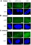
Representative immunofluorescence images of pATM foci and pATM crowns (green) were observed in the indicated cells at the 1st week (HLEpic and GM03399) or the 3rd (EROS08O) week of exposure to 120 mSv/y and of HDC at the (A) 1st (HLEpic), (B) 3rd (EROS08O), and (C) 4th week (GM033999). Each image has been counterstained with DAPI (blue). The white bar represents 5 µm.
Figure 5
Figure 5
Hypothetic model of formation of pATM crowns and HDC. When permanent stress is applied to cells showing a perinuclear overexpression of an X-protein (A), progressively, the ATM monomers bind to X-proteins. At this step, since ATM monomers can enter the nucleus, DSB recognition and repair are still possible (B). When the first layer of the pATM crown is complete, DSB recognition and repair are impossible (C). Since ATM monomers cumulate around the nucleus, such a high concentration of ATM monomers leads to the re-dimerization of ATM (D). The DNA strand breaks cumulate in the nucleus, and the chromatin decondenses and enters some ATM monomers that recognize the numerous DSB by forming γH2AX and ATM foci: the cell becomes HDC (E).
Figure 6
Figure 6
Theoretical illustration of the ATM crowns and HDC signals as a function of time. The occurrence of ATM crowns (in black) systematically precedes the formation of HDC (in red). However, the measurement may reveal much more or much less HDC than ATM crowns (the dotted or dashed lines reflect different response in the formation of HDC.
Figure 7
Figure 7
Schematic representation of the protein interaction described by our mathematical model. In the cytoplasm (light green background), ATM dimers are monomerized by oxidative stress, and ATM monomers migrate near the nucleus (step 1 in the figure). ATM monomers may eventually cross the nucleus membrane and reach the nucleus (light blue background) to detect DSB and trigger their repair process (step 2). On their way to the nucleus, cytoplasmic ATM monomers may form ATM-X protein complexes as well as ATM dimers around the nucleus, leading to the formation of a perinuclear ATM crown (light red background) (step 3). When the perinuclear ATM crown is formed and thick enough, cytoplasmic ATM monomers cannot reach the nucleus anymore.
Figure 8
Figure 8
Numerical simulation representing the qualitative behavior of the different protein concentrations as a function of time. (A) We observed that as the perinuclear ATM crown (due to forced ATM concentration, in dashed blue curve) reaches a certain threshold (two weeks here), the radius of the nucleus (in blue curve) increases up to 3-folds its initial size, leading to the release of the induced stress (in orange curve). (B) These events lead to a monomerization of the ATM dimers (ATM dimers and ATM-X-protein–ATM complexes) in the crown (green and magenta curves). These events lead to the destruction of the pATM crown. In addition, the newly formed cytoplasmic ATM monomers may cross the nucleus and recognize the DSB (black curve).

References

    1. Cucinotta F.A., Kim M.-H.Y., Ren L. Managing Luna and Mars Mission Radiation Risks. Part I; Cancer Risks, Uncertainties and Shielding Effectiveness. NASA; Hanover, MD, USA: 2005. NASA Report. TP-2005-213164.
    1. Durante M., Cucinotta F.A. Heavy ion carcinogenesis and human space exploration. Nat. Rev. Cancer. 2008;8:465–472. doi: 10.1038/nrc2391. - DOI - PubMed
    1. Durante M. New challenges in high-energy particle radiobiology. Br. J. Radiol. 2014;87:20130626. doi: 10.1259/bjr.20130626. - DOI - PMC - PubMed
    1. Cucinotta F.A., Schimmerling W., Wilson J.W., Peterson L.E., Badhmar G.D., Saganti P.B., Dicello J.F. Space Radiation Cancer Risk Projections for Exploration Missions: Uncertainty Reduction and Mitigation. NASA; Hanover, MD, USA: 2011. NASA Report. JSC-29295.
    1. Maalouf M., Durante M., Foray N. Biological effects of space radiation on human cells: History, advances and outcomes. J. Radiat. Res. 2011;52:126–146. doi: 10.1269/jrr.10128. - DOI - PubMed

Publication types

MeSH terms

Substances

LinkOut - more resources