. 2024 Oct 15;13(20):1704.
doi: 10.3390/cells13201704.

Decreased Circulating Gonadotropin-Releasing Hormone Associated with Keratoconus

Affiliations

Decreased Circulating Gonadotropin-Releasing Hormone Associated with Keratoconus

Paulina Escandon et al. Cells. .

Abstract

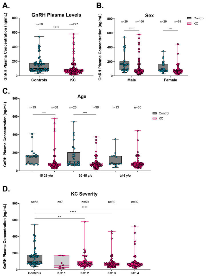
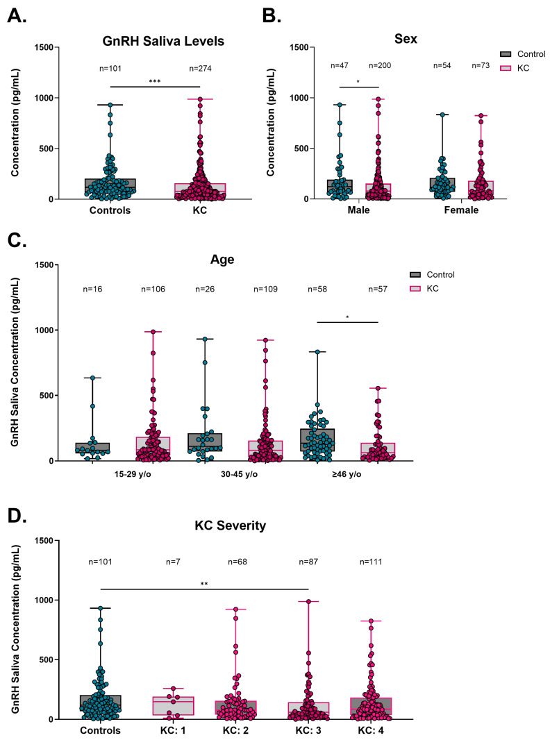
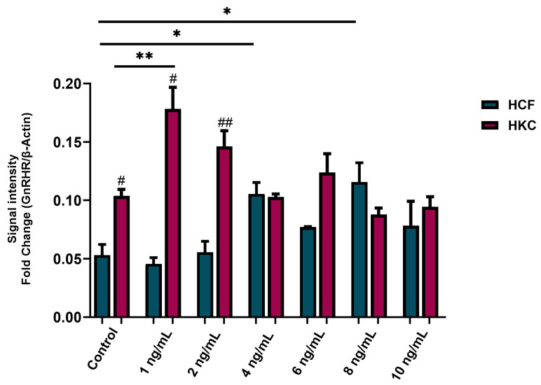
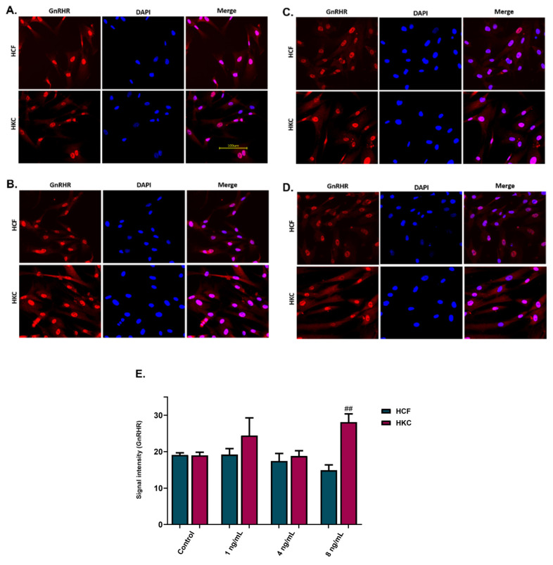
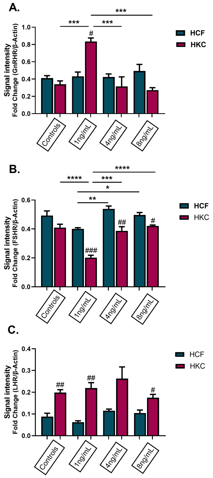
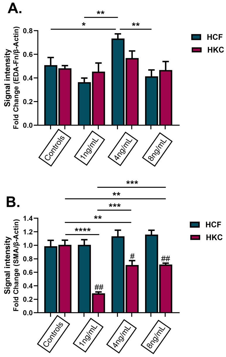
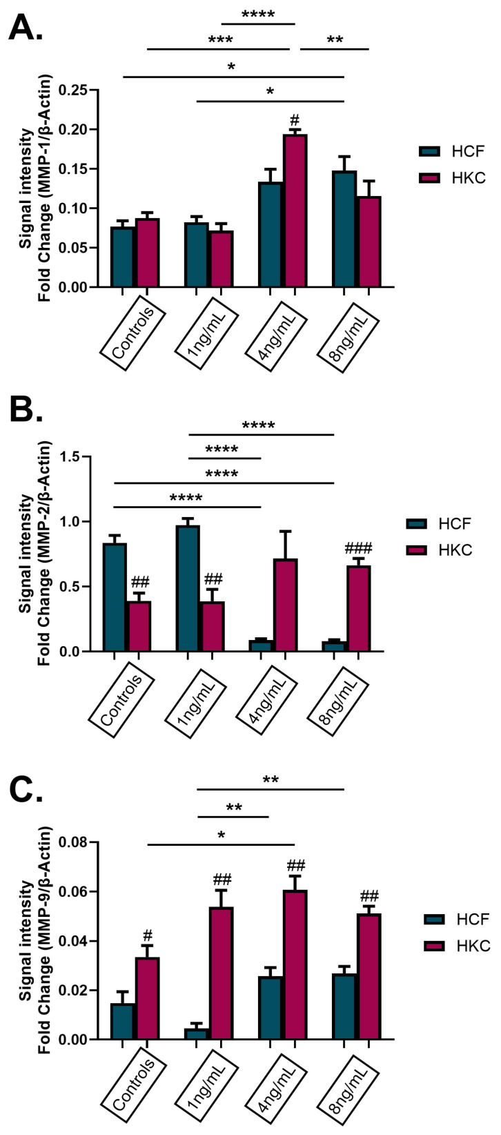
Keratoconus (KC) is a corneal thinning dystrophy that leads to visual impairment. While the cause of KC remains poorly understood, changes in sex hormone levels have been correlated with KC development. This study investigated circulating gonadotropin-releasing hormone (GnRH) in control and KC subjects to determine if this master hormone regulator is linked to the KC pathology. Plasma and saliva were collected from KC subjects (n = 227 and n = 274, respectively) and non-KC controls (n = 58 and n = 101, respectively), in concert with patient demographics and clinical features. GnRH levels in both plasma and saliva were significantly lower in KC subjects compared to controls. This finding was retained in plasma when subjects were stratified based on age, sex, and KC severity. Control and KC corneal fibroblasts (HKCs) stimulated with recombinant GnRH protein in vitro revealed significantly increased luteinizing hormone receptor by HKCs and reduced expression of α-smooth muscle actin with treatment suggesting that GnRH may modulate hormonal and fibrotic responses in the KC corneal stroma. Further studies are needed to reveal the role of the hypothalamic-pituitary-gonadal axis in the onset and progression of KC and to explore this pathway as a novel therapeutic target.

Keywords: biomarkers; corneal thinning; crosslinking; estrogen; keratoconus; saliva.


Conflict of interest statement

The authors declare no conflicts of interest.

Figures

Figure 1
Figure 1
Plasma gonadotropin-releasing hormone (GnRH) in control and KC patients. (A) GnRH concentration in plasma for all control and KC patients tested. Sub-analyses of GnRH concentrations based on (B) sex, (C) age, and (D) KC severity. Data are shown in box-and-whisker plots, with the box represent the 25% quartile, median, and 75% quartile, and the line extending to the minimum and maximum values. Statistical significance evaluated by a non-parametric Mann–Whitney test (AC). Statistical significance evaluated by a non-parametric Kruskal–Wallis test with Dunn’s multiple comparisons test (D). Significance indicated as ** p < 0.01, *** p < 0.001, and **** p < 0.0001.
Figure 2
Figure 2
Saliva gonadotropin-releasing hormone (GnRH) in control and KC patients. (A) GnRH concentration in saliva for all control and KC patients tested. Sub-analyses of GnRH concentrations based on (B) sex, (C) age, and (D) KC severity. Data are shown in box-and-whisker plots, with the box representing the 25% quartile, median, and 75% quartile, and the line extending to the minimum and maximum values. Statistical significance evaluated by a non-parametric Mann–Whitney test (AC). Statistical significance evaluated by a non-parametric Kruskal–Wallis test with Dunn’s multiple comparisons test (D). Significance indicated as * p < 0.05, ** p < 0.01, and *** p < 0.001.
Figure 3
Figure 3
Protein expression of gonadotropin-releasing hormone receptor (GnRHR) in 2D HCFs and HKCs after being stimulated with 1 ng/mL, 2 ng/mL, 4 ng/mL, 6 ng/mL, 8 ng/mL, and 10 ng/mL rGnRH for 48 h. Data shown as mean ± standard error of the mean. n = 3 per group. Statistical significance of dose with HCFs and HKCs based on a one-way ANOVA with Šídák’s multiple comparisons test. Post hoc significance indicated as * p < 0.05 and ** p < 0.01. Statistical significance between HCFs and HKCs within dose determined by unpaired t-test with Welch’s correction. Significance indicated as # p < 0.05, ## p < 0.01.
Figure 4
Figure 4
Immunofluorescence images and analysis for gonadotropin-releasing hormone receptor (GnRHR) expression in 2D GnRH stimulation of HCFs and HKCs. (A) The controls were not stimulated with GnRH; (B) 1 ng/mL; (C) 4 ng/mL; (D) 8 ng/mL; (E) immunofluorescence images’ signal intensity for GnRHR expression. Statistical significance between HCFs and HKCs within the dose determined by an unpaired t-test with Welch’s correction. Significance indicated as ## p < 0.01.
Figure 5
Figure 5
Protein expression of hormone receptors in 3D HCFs and HKCs following stimulation with 1 ng/mL, 4 ng/mL, and 8 ng/mL GnRH for 4 weeks. Protein expression of (A) GnRH receptor, (B) FSH receptor, and (C) LH receptor based on Western blot analysis. Statistical significance based on a one-way ANOVA with Šídák’s multiple comparisons test. Post hoc significance indicated as * p < 0.05, ** p < 0.01, *** p < 0.001, and **** p < 0.0001. Statistical significance between HCFs and HKCs within the dose determined by an unpaired t-test with Welch’s correction. Significance indicated as # p < 0.05, ## p < 0.01, and ### p < 0.001.
Figure 6
Figure 6
Protein expression of fibronectin and α-smooth muscle actin (SMA) in 3D HCFs and HKCs following stimulation with 1 ng/mL, 4 ng/mL, and 8 ng/mL GnRH for 4 weeks. (A) Results of EDA-Fn protein expression in HCFs and HKCs stimulated with GnRH. (B) Results of SMA protein expression in HCFs and HKCs stimulated with GnRH. Statistical significance based on a one-way ANOVA with Šídák’s multiple comparisons test. Post hoc significance indicated as * p < 0.05, ** p < 0.01, *** p < 0.001, **** p < 0.0001. Statistical significance between HCFs and HKCs within the dose determined by the unpaired t-test with Welch’s correction. Significance indicated as # p < 0.05, ## p < 0.01.
Figure 7
Figure 7
Protein expression of matrix metalloproteinases (MMPs) in 3D HCFs and HKCs after being stimulated with 1 ng/mL, 4 ng/mL, and 8 ng/mL GnRH for 4 weeks. Protein expression of (A) MMP-1, (B) MMP-2, and (C) MMP-9 in HCFs and HKCs stimulated with GnRH. Statistical significance based on a one-way ANOVA with Šídák’s multiple comparisons test. Post hoc significance indicated as * p < 0.05, ** p < 0.01, *** p < 0.001, and **** p < 0.0001. Statistical significance between HCFs and HKCs within the dose determined by the unpaired t-test with Welch’s correction. Significance indicated as # p < 0.05, ## p < 0.01, and ### p < 0.001.

References

    1. Kennedy R.H., Bourne W.M., Dyer J.A. A 48-year clinical and epidemiologic study of keratoconus. Am. J. Ophthalmol. 1986;101:267–273. doi: 10.1016/0002-9394(86)90817-2. - DOI - PubMed
    1. Godefrooij D.A., de Wit G.A., Uiterwaal C.S., Imhof S.M., Wisse R.P. Age-specific Incidence and Prevalence of Keratoconus: A Nationwide Registration Study. Am. J. Ophthalmol. 2017;175:169–172. doi: 10.1016/j.ajo.2016.12.015. - DOI - PubMed
    1. Papali’i-Curtin A.T., Cox R., Ma T., Woods L., Covello A., Hall R.C. Keratoconus Prevalence Among High School Students in New Zealand. Cornea. 2019;38:1382–1389. doi: 10.1097/ICO.0000000000002054. - DOI - PubMed
    1. Galgauskas S., Juodkaite G., Tutkuvienė J. Age-related changes in central corneal thickness in normal eyes among the adult Lithuanian population. Clin. Interv. Aging. 2014;9:1145–1151. doi: 10.2147/CIA.S61790. - DOI - PMC - PubMed
    1. Chatzis N., Hafezi F. Progression of keratoconus and efficacy of pediatric [corrected] corneal collagen cross-linking in children and adolescents. J. Refract. Surg. 2012;28:753–758. doi: 10.3928/1081597X-20121011-01. - DOI - PubMed

Publication types

Substances

LinkOut - more resources