. 2024 Oct 17;13(20):1715.
doi: 10.3390/cells13201715.

Difenoconazole Induced Damage of Bovine Mammary Epithelial Cells via ER Stress and Inflammatory Response

Affiliations

Difenoconazole Induced Damage of Bovine Mammary Epithelial Cells via ER Stress and Inflammatory Response

Myoung-Jun Na et al. Cells. .

Abstract

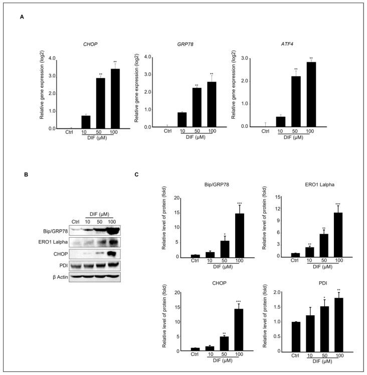
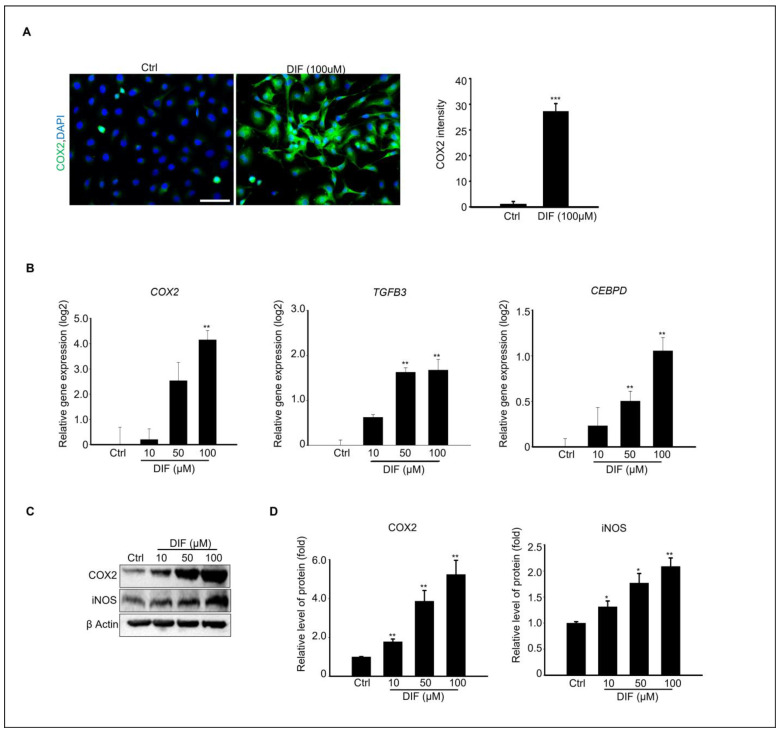
Difenoconazole (DIF) is a fungicide used to control various fungi. It is absorbed on the surface of different plants and contributes significantly to increased crop production. However, DIF is reported to exhibit toxicity to fungi and to aquatic plants, fish, and mammals, including humans, causing adverse effects. However, research on the impact of DIF on the mammary epithelial cells of herbivorous bovines is limited. DIF-induced damage and accumulation in the mammary glands can have direct and indirect effects on humans. Therefore, we investigated the effects and mechanisms of DIF toxicity in MAC-T cells. The current study revealed that DIF reduces cell viability and proliferation while triggering apoptotic cell death through the upregulation of pro-apoptotic proteins, including cleaved caspase 3 and Bcl-2-associated X protein (BAX), and the downregulation of leukemia type 2 (BCL-2). DIF also induced endoplasmic reticulum (ER) stress by increasing the expression of genes or proteins of Bip/GRP78, protein disulfide isomerase (PDI), activating transcription factor 4 (ATF4), C/EBP homologous protein (CHOP), and endoplasmic reticulum oxidoreductase 1 Alpha (ERO1-Lα). We demonstrated that DIF induces mitochondria-mediated apoptosis in MAC-T cells by activating ER stress pathways. This cellular damage resulted in a significant increase in the expression of inflammatory response genes and proteins, including cyclooxygenase 2 (COX2), transforming growth factor beta 3 (TGFB3), CCAAT enhancer binding protein delta (CEBPD), and iNOS, in DIF-treated groups. In addition, spheroid formation by MAC-T cells was suppressed by DIF treatment. Our findings suggest that DIF exposure in dairy cows may harm mammary gland function and health and may indirectly affect human consumption of milk.

Keywords: ER stress; bovine mammary gland; difenoconazole; inflammation.


Conflict of interest statement

The authors declare that they do not have any conflicts of interests.

Figures

Figure 1
Figure 1
Effects of DIF on MAC-T cells include significant impacts on both cell apoptosis and proliferation. (A) MAC-T cell viability through an MTT assay. Cells were treated with DIF (0–1000 µM). Data represent the mean ± SD (n = 5, ** p < 0.01, *** p < 0.001 compared to the control). (B) After treating cells with DIF (0–100 μM) followed by a 24 h incubation period, the morphology of the cells was observed using a microscope, and immunostaining was performed using Ki-67 antibody. Scaler bar = 100 μM. (C) The graph illustrating the ratio of Ki-67-positive cells to the total cells stained with DAPI has been created for the manuscript (n = 4, ** p < 0.001). (D) After culturing MAC-T cells with DIF at concentrations ranging from 0 to 100 μM, annexin V-FITC/PI staining was conducted. Following staining, dye-positive/negative cells were assessed using flow cytometry to determine cell death by apoptosis. (E) The graph depicts the proportion of apoptosis as mean ± SD based on the results of flow cytometry analysis. (n = 4, ** p < 0.001).
Figure 2
Figure 2
Expression levels of Pro-apoptotic protein, and mitochondrial dysfunction in DIF exposed MAC-T cell. (A) MAC-T cells were treated with DIF concentrations ranging from 0 to 100 μM for 24 h, followed by preparation of total protein and analysis via immunoblotting. The protein expression levels of cleaved caspase 3, caspase 3 and BAX, Bcl-2, and β-actin were evaluated in each experimental group. (B) The graphs display protein bands represented as quantified data normalized to β-actin as mean ± SD (n = 5, * p < 0.05, ** p < 0.01, *** p < 0.001). (C) Cells treated with DIF (0–100 μM) were cultured in a 6-well plate, followed by JC-1 staining. Red and green fluorescence were observed using fluorescence microscopy. The ratio of JC-1 green emitted at 530 nm and JC-1 red emitted at 580 nm varies depending on the change in mitochondrial membrane potential. Scale bar =100 μm. (D) Flow cytometry analysis was conducted for quantification of JC-1 green and red. (E) A graph was generated based on the decrease in the proportion predicted to be JC-1 red fluorescence (% J-monomers/J-aggregates), and data show mean ± SD. (n = 5, ** p < 0.01, and *** p < 0.001).
Figure 3
Figure 3
Effects of DIF on ER stress in MAC-T cell. (A) qPCR analysis of CHOP, Bip/Grp78, and ATF4 expression levels following 24 h of DIF treatment. Data represent mean ± SD with log2 scales (n = 5, ** p < 0.01 compared to controls). (B) Immunoblot analysis showing the protein levels of BiP/GRP78, ERO1-Lα, CHOP, PDI, and β-actin after DIF exposure. (C) Densitometric quantification of protein bands normalized to β-actin. Results are presented as the mean ± SD (n = 5, * p < 0.05, ** p < 0.01, *** p < 0.001).
Figure 4
Figure 4
DIF induced inflammation at cultured MAC-T cell. (A) Immunostaining of COX2 protein in100 μM DIF treated MAC-T cells. Scale bar = 50 μM. DAPI were stained nucleus. Graph showed COX2 intensity from each group. *** p < 0.001, (B) The relative gene expression of Cox2, TGFB2, and CEBPD in each group after 0–100 μM DIF treatment for 24 h. Graph showed mean ± SD (n = 5, ** p < 0.001 compared to controls). (C) Protein expression of COX2, iNOS, and β-actin after 0–100 μM DIF exposure. (D) Graph represents each protein band normalized to β-actin as mean ± SD (n = 5, * p < 0.05, ** p < 0.01 compared to controls).
Figure 5
Figure 5
Effects of DIF on spheroid formation from MAC-T cells. (A) Morphological change in spheroid from MAC-T cell after 0–100 μM DIF treatment for 3 d. Images were captured using bright-field microscopy, with the scale bar representing = 500 μM. (B) GFP expressing spheroid of MAC-T cell (green fluorescent) after 0–100 DIF μM treatment. Scale bar = 500 μM. (C) The area of spheroid (%) from each group. Graph showed mean ± SD (** p < 0.01, *** p < 0.001 compared to controls). Spheroid formation did not occur in 100 μM DIF-treated groups. (D) Average diameter (μm) of spheroid from each group. The diameter measurement was carried out from D1–D3. Graph showed mean ± SD (* p < 0.05, ** p < 0.01, *** p < 0.001 compared to controls).

Similar articles

Cited by

References

    1. Machado F.J., Santana F.M., Lau D., Del Ponte E.M. Quantitative Review of the Effects of Triazole and Benzimidazole Fungicides on Fusarium Head Blight and Wheat Yield in Brazil. Plant Dis. 2017;101:1633–1641. doi: 10.1094/PDIS-03-17-0340-RE. - DOI - PubMed
    1. Rather I.A., Sabir J.S.M., Asseri A.H., Wani M.Y., Ahmad A. Triazole Derivatives Target 14α-Demethylase (LDM) Enzyme in Candida albicans Causing Ergosterol Biosynthesis Inhibition. J. Fungi. 2022;8:688. doi: 10.3390/jof8070688. - DOI - PMC - PubMed
    1. Brauer V.S., Rezende C.P., Pessoni A.M., De Paula R.G., Rangappa K.S., Nayaka S.C., Gupta V.K., Almeida F. Antifungal Agents in Agriculture: Friends and Foes of Public Health. Biomolecules. 2019;9:521. doi: 10.3390/biom9100521. - DOI - PMC - PubMed
    1. Schäfer R.B., Pettigrove V., Rose G., Allinson G., Wightwick A., von der Ohe P.C., Shimeta J., Kühne R., Kefford B.J. Effects of pesticides monitored with three sampling methods in 24 sites on macroinvertebrates and microorganisms. Environ. Sci. Technol. 2011;45:1665–1672. doi: 10.1021/es103227q. - DOI - PubMed
    1. Luong T.T., Nguyen T.H.T., Nguyen T.D., Le V.T., Pham T.H.T., Ho T.T., Nguyen N.L. Degradation of Triazole Fungicides by Plant Growth-Promoting Bacteria from Contaminated Agricultural Soil. J. Microbiol. Biotechnol. 2024;34:56–64. doi: 10.4014/jmb.2308.08037. - DOI - PMC - PubMed

Publication types

LinkOut - more resources