. 2024 Oct 18;13(20):1731.
doi: 10.3390/cells13201731.

Multispectral Imaging of Collagen, NAD(P)H and Flavin Autofluorescence in Mesenchymal Stem Cells Undergoing Trilineage Differentiation

Affiliations

Multispectral Imaging of Collagen, NAD(P)H and Flavin Autofluorescence in Mesenchymal Stem Cells Undergoing Trilineage Differentiation

Jared M Campbell et al. Cells. .

Abstract

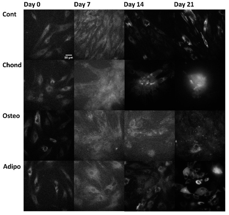
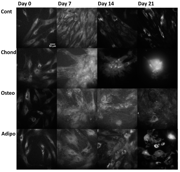
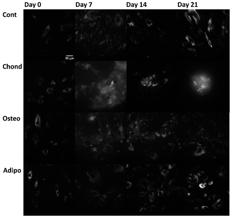
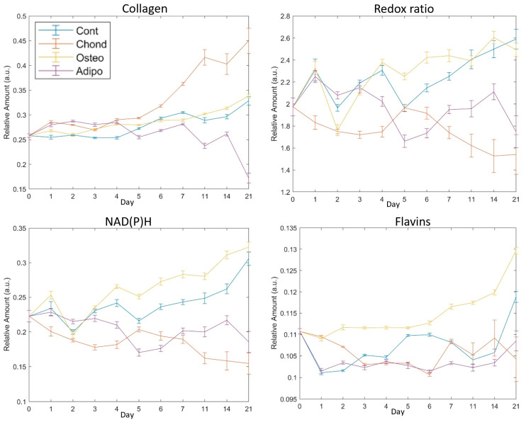
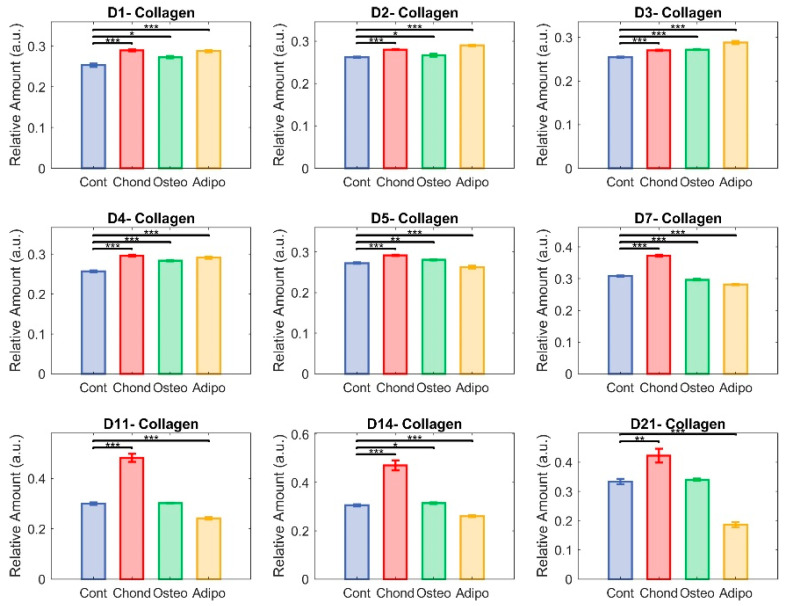
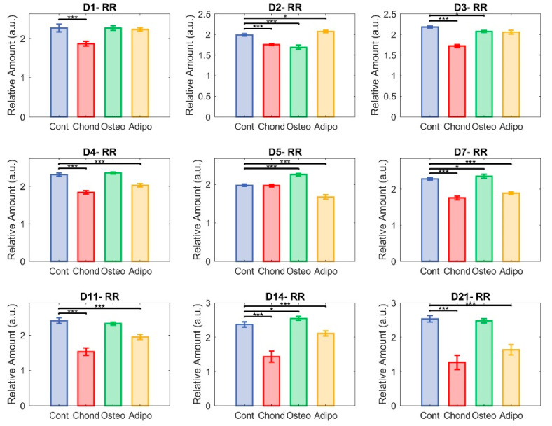
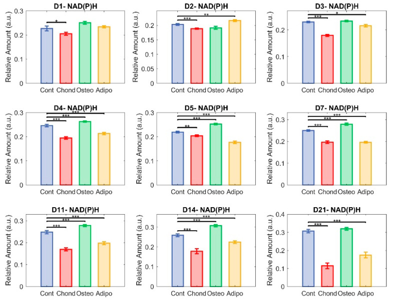
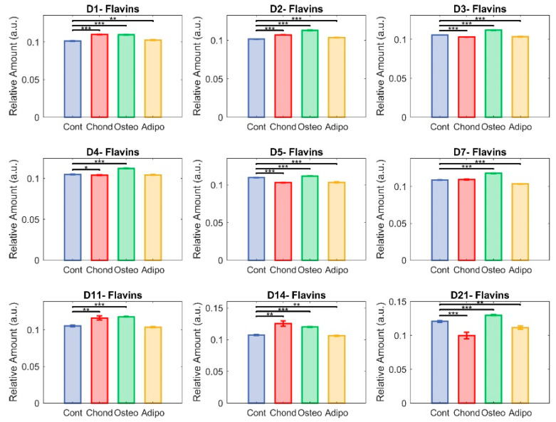
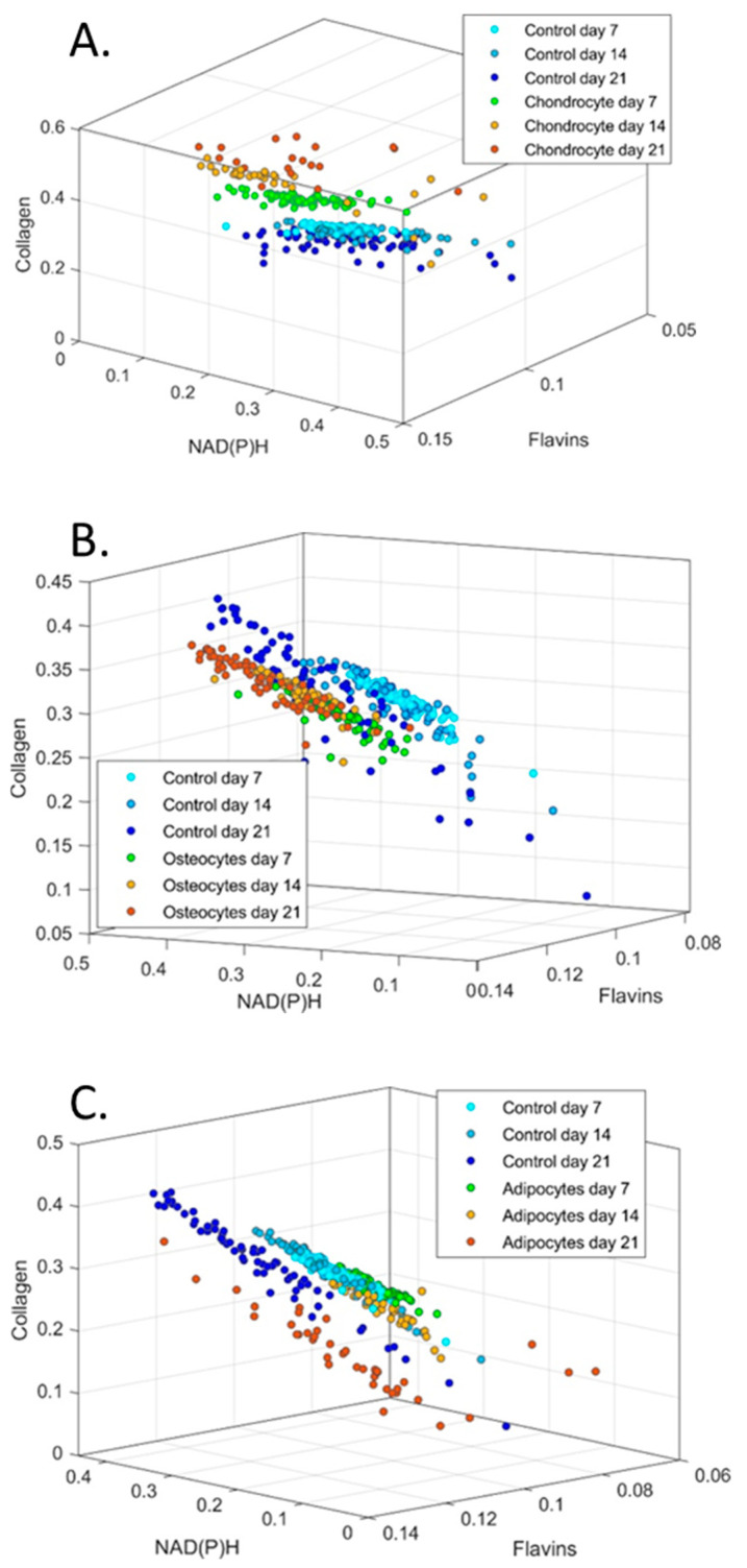
Understanding the molecular mechanisms of differentiation is important for regenerative medicine and developmental biology. This study aims to characterise the role of the glycolysis/oxidative phosphorylation balance as a driver of mesenchymal stem cell (MSC) differentiation. Cells were maintained in normal conditions or stimulated towards the MSC trilineage cell types over 21 days. Multispectral imaging of cell autofluorescence was applied as a non-invasive methodology to continuously image cultures in situ. Spectral signals for collagen, NAD(P)H, and flavins were unmixed. MSCs cultured under chondrogenic conditions exhibited increased collagen levels relative to controls. Following osteogenic induction, MSCs showed increased collagen levels relative to controls during the earlier stages of culture; however, control cells increased their collagen levels as they became confluent. MSCs cultured under adipogenic conditions exhibited lower levels of collagen than controls. The redox ratio (RR; NAD(P)H/flavins) immediately decreased during chondrogenesis, with this early effect persisting throughout the culture compared to control cells, which appeared to increase their RR, similar to osteogenesis. Adipogenesis resulted in a small increase in RR on day 2 relative to control cells, followed by a persistent decrease. Chondrogenic and adipogenic differentiation favoured oxidative phosphorylation, whereas osteogenesis and MSC overgrowth resulted in a glycolytic metabolism. Following consideration of these findings, as well as the diverse reports in the literature, it is concluded that neither enhanced oxidative phosphorylation nor glycolysis are fundamental to the canonical modes of differentiation, and researchers should avoid interpreting shifts as indicating differentiation.

Keywords: autofluorescence; differentiation; mesenchymal; spectroscopy; stem cells.


Conflict of interest statement

The authors report no conflicts of interest.

Figures

Figure 1
Figure 1
Cultured MSCs stimulated towards (A) chondrogenesis, (B) osteogenesis, (C) adipogenesis, all stained with safranin-O, alizarin red, and oil red O, respectively, on day 21 of culture.
Figure 2
Figure 2
Collagen (not specific to any type) autofluorescence (arising from the excitation at 371 nm and emission at 414 nm, channel where collagen is the dominant—although not the sole—autofluorophore) from control, chondrogenic, osteogenic, and adipogenic cells on days 0, 7, 14, and 21.
Figure 3
Figure 3
NAD(P)H autofluorescence, arising from the excitation at 345 nm and emission at 451 nm (where NAD(P)H is the dominant—although not the sole—autofluorophore), from control, chondrogenic, osteogenic, and adipogenic cells on days 0, 7, 14, and 21.
Figure 4
Figure 4
Flavin autofluorescence, arising from the excitation at 490 nm and emission at 575 nm (where flavin proteins are the dominant—although not the sole—autofluorophores), from control, chondrogenic, osteogenic, and adipogenic cells on days 0, 7, 14, and 21.
Figure 5
Figure 5
Mean values of fluorophores with SEM error bars on each assessed day for collagen and the redox ratio of NAD(P)H to flavins. Day 0 indicates cells with no exposure to the differentiation media, day 21 was the final assessment day. The redox ratio is NAD(P)H/flavins.
Figure 6
Figure 6
Unmixed collagen. Here, each group—control (in blue), chondrocytes (in red), osteocytes (in green), and adipocytes (in yellow)—is represented with respect to their relative content of collagen. The non-parametric Mann–Whitney U test was used to assess significance (* p < 0.05, ** p < 0.01, and *** p < 0.001) among groups.
Figure 7
Figure 7
Redox ratio (RR). Each group—control (in blue), chondrocytes (in red), osteocytes (in green), and adipocytes (in yellow)—represent NAD(P)H relative abundance divided by flavin relative abundance in the cultured cells (* p < 0.05 and *** p < 0.001).
Figure 8
Figure 8
Unmixed NAD(P)H. Here, each group—control (in blue), chondrocytes (in red), osteocytes (in green), and adipocytes (in yellow)—is represented in terms of the relative abundance of NAD(P)H (* p < 0.05, ** p < 0.01, and *** p < 0.001).
Figure 9
Figure 9
Unmixed flavins. Here, each group—control (in blue), chondrocytes (in red), osteocytes (in green), and adipocytes (in yellow)—is represented in terms of relative abundance of flavins (* p < 0.05, ** p < 0.01, and *** p < 0.001).
Figure 10
Figure 10
Cluster graphs of relative abundance of collagen, NAD(P)H and flavins plotted against each other for (A) chondrocytes, (B) osteocytes and (C) adipocytes. 3D graphs have been oriented to best show groupings. Control cells are plotted in each group for comparison.

Similar articles

References

    1. Pittenger M.F., Discher D.E., Peault B.M., Phinney D.G., Hare J.M., Caplan A.I. Mesenchymal stem cell perspective: Cell biology to clinical progress. NPJ Regen. Med. 2019;4:22. doi: 10.1038/s41536-019-0083-6. - DOI - PMC - PubMed
    1. Giwa R., Brestoff J.R. Mitochondria Transfer to CD4+ T Cells May Alleviate Rheumatoid Arthritis by Suppressing Pro-Inflammatory Cytokine Production. Immunometabolism. 2022;4:e220009. doi: 10.20900/immunometab20220009. - DOI - PMC - PubMed
    1. Islam M.N., Das S.R., Emin M.T., Wei M., Sun L., Westphalen K., Rowlands D.J., Quadri S.K., Bhattacharya S., Bhattacharya J. Mitochondrial transfer from bone-marrow-derived stromal cells to pulmonary alveoli protects against acute lung injury. Nat. Med. 2012;18:759–765. doi: 10.1038/nm.2736. - DOI - PMC - PubMed
    1. Varum S., Rodrigues A.S., Moura M.B., Momcilovic O., Easley C.A.t., Ramalho-Santos J., Van Houten B., Schatten G. Energy metabolism in human pluripotent stem cells and their differentiated counterparts. PLoS ONE. 2011;6:e20914. doi: 10.1371/journal.pone.0020914. - DOI - PMC - PubMed
    1. Meleshina A.V., Dudenkova V.V., Bystrova A.S., Kuznetsova D.S., Shirmanova M.V., Zagaynova E.V. Two-photon FLIM of NAD(P)H and FAD in mesenchymal stem cells undergoing either osteogenic or chondrogenic differentiation. Stem Cell Res. Ther. 2017;8:15. doi: 10.1186/s13287-017-0484-7. - DOI - PMC - PubMed

Publication types

LinkOut - more resources