Review
. 2024 Oct 20;13(20):1738.
doi: 10.3390/cells13201738.

Apolipoprotein-L1 (APOL1): From Sleeping Sickness to Kidney Disease

Affiliations
Review

Apolipoprotein-L1 (APOL1): From Sleeping Sickness to Kidney Disease

Etienne Pays. Cells. .

Abstract

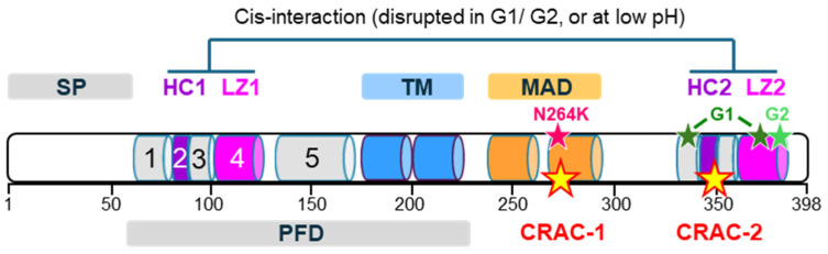
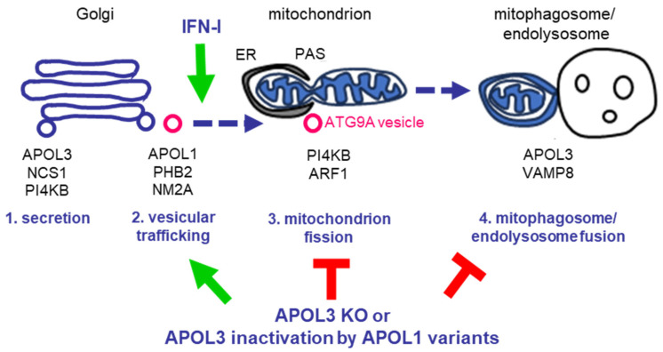
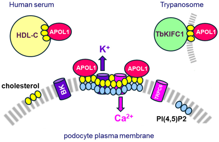
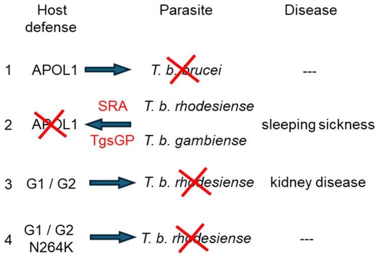
Apolipoprotein-L1 (APOL1) is a membrane-interacting protein induced by inflammation, which confers human resistance to infection by African trypanosomes. APOL1 kills Trypanosoma brucei through induction of apoptotic-like parasite death, but two T. brucei clones acquired resistance to APOL1, allowing them to cause sleeping sickness. An APOL1 C-terminal sequence alteration, such as occurs in natural West African variants G1 and G2, restored human resistance to these clones. However, APOL1 unfolding induced by G1 or G2 mutations enhances protein hydrophobicity, resulting in kidney podocyte dysfunctions affecting renal filtration. The mechanism involved in these dysfunctions is debated. The ability of APOL1 to generate ion pores in trypanosome intracellular membranes or in synthetic membranes was provided as an explanation. However, transmembrane insertion of APOL1 strictly depends on acidic conditions, and podocyte cytopathology mainly results from secreted APOL1 activity on the plasma membrane, which occurs under non-acidic conditions. In this review, I argue that besides inactivation of APOL3 functions in membrane dynamics (fission and fusion), APOL1 variants induce inflammation-linked podocyte toxicity not through pore formation, but through plasma membrane disturbance resulting from increased interaction with cholesterol, which enhances cation channels activity. A natural mutation in the membrane-interacting domain (N264K) abrogates variant APOL1 toxicity at the expense of slightly increased sensitivity to trypanosomes, further illustrating the continuous mutual adaptation between host and parasite.

Keywords: APOL1 risk variants; ARF1; INF-I inflammation; N264K variant; NCS1; PI4KB; apoptosis; cation channels; cholesterol microdomains; chronic kidney disease; membrane dynamics; mitochondrion fission; mitophagy; sleeping sickness.


Conflict of interest statement

The author declares no conflicts of interest.

Figures

Figure 1
Figure 1
Scheme of APOL1 structure (398 amino acids). The different colored cylinders are different α-helices. SP = signal peptide; HC = hydrophobic cluster; LZ = leucine zipper; TM = transmembrane span; MAD = membrane-addressing domain; PFD = pore-forming domain; CRAC = cholesterol recognition amino acid consensus. The approximate locations of the N264K, G1, and G2 mutations are indicated by pink, dark green, and light green stars, respectively, and the CRAC motifs are indicated by yellow stars.
Figure 2
Figure 2
APOL1 involvement in membrane dynamics [26,27,44]. Under non-inflammatory conditions (1), APOL1 is associated with APOL3 at trans-Golgi and endoplasmic reticulum membranes. APOL3 controls PI4KB activity for vesicular secretion in a trimeric complex with NCS1. Upon infection-induced inflammation (2), IFN-I signaling triggers ARF1 activation and dissociation of PI4KB from the Golgi in Golgi-derived ATG9A vesicles also carrying APOL1, APOL3, PHB2, and NM2A. While NM2A drives vesicular trafficking, the mitophagy receptor PHB2 targets the vesicles to mitochondrial pre-autophagosomal structures (PAS) at mitochondrion–endoplasmic reticulum (ER) contact sites. PI4KB and ARF1 induce mitochondrial membrane fission, releasing mitophagosomes (3). The mitophagosome fusion with endolysosomes (4) allows completion of mitophagy. This fusion is triggered by interactions between APOL3 and endosomal VAMP8. At each step, relevant key protein complexes are mentioned. Either APOL3 KO or APOL3 inactivation by APOL1 C-terminal variants leads to PI4KB dissociation from the Golgi in a process mimicking that induced by IFN-I, except that the absence of ARF1 activation and lack of APOL3 activity result in defective mitophagy (incomplete membrane fission and fusion). In addition to the induction of mitophagy, APOL1 trafficking activity to the mitochondrion also conditions APOL3-mediated apoptosis induced under inflammatory conditions.
Figure 3
Figure 3
The role of cholesterol in APOL1 trafficking and toxic activity (adapted from [25]). APOL1 is associated with the serum cholesterol carrier HDL-C or with the T. brucei cholesterol carrier TbKIFC1. At the surface of kidney podocytes, the activity of Ca2+ TRPC6 channels and Ca2+-dependent K+ BK channels is controlled in cholesterol microdomains involving the cholesterol-binding podocin and the actin cytoskeleton. Through interaction with cholesterol in the upper layer of the podocyte plasma membrane [25], APOL1 variants may increase cation fluxes by TRPC6 and BK channels, causing cytotoxicity. Normally, secreted APOL1 is sequestered by HDL-C particles, restraining the availability of APOL1 for interaction with the podocyte surface. However, such limitation is lost under two types of situations: in cell culture in vitro, or upon inflammation in vivo, such occurs upon virus infection. In both cases, APOL1 synthesis is increased whereas the HDL-C amount is reduced [3,4,88,89,90], increasing APOL1 availability for toxic surface interaction. Accordingly, low HDL cholesterol is a known predictor of chronic kidney disease [91,92,93,94].
Figure 4
Figure 4
Molecular ‘arms race’ between African trypanosomes and humans. (1) Human ancestors produced a secreted version of APOL1 to kill the extracellular parasite Trypanosoma brucei brucei. (2) Two T. b. brucei clones, known as T. b. rhodesiense and T. b. gambiense, used variant surface glycoprotein (VSG)-derived proteins termed SRA and TgsGP, respectively, to neutralize APOL1. (3) The APOL1 C-terminal variants G1 and G2 allowed human resistance to SRA, hence human ability to kill T. b. rhodesiense. However, this also resulted in a high probability of humans developing chronic kidney disease. (4) C-terminal APOL1 variants with the N264K mutation can still efficiently lyze T. b. rhodesiense, but no longer exhibit kidney cytotoxicity.

Similar articles

Cited by

References

    1. Pays E. The function of Apolipoproteins L (APOLs): Relevance for kidney disease, neurotransmission disorders, cancer and viral infection. FEBS J. 2021;288:360–381. doi: 10.1111/febs.15444. - DOI - PMC - PubMed
    1. Duchateau P.N., Pullinger C.R., Orellana R.E., Kunitake S.T., Naya-Vigne J., O’Connor P.M., Malloy M.J., Kane J.P. Apolipoprotein L, a new human high density lipoprotein apolipoprotein expressed by the pancreas. Identification, cloning, characterization, and plasma distribution of apolipoprotein L. J. Biol. Chem. 1997;272:25576–25582. doi: 10.1074/jbc.272.41.25576. - DOI - PubMed
    1. Nichols B., Jog P., Lee J.H., Blackler D., Wilmot M., D’Agati V., Markowitz G., Kopp J.B., Alper S.L., Pollak M.R., et al. Innate immunity pathways regulate the nephropathy gene Apolipoprotein L1. Kidney Int. 2015;87:332–342. doi: 10.1038/ki.2014.270. - DOI - PMC - PubMed
    1. Uzureau S., Coquerelle C., Vermeiren C., Uzureau P., Van Acker A., Pilotte L., Monteyne D., Acolty V., Vanhollebeke B., Van den Eynde B., et al. Apolipoproteins L control cell death triggered by TLR3/TRIF signaling in dendritic cells. Eur. J. Immunol. 2016;46:1854–1866. doi: 10.1002/eji.201546252. - DOI - PubMed
    1. Smith E.E., Malik H.S. The apolipoprotein L family of programmed cell death and immunity genes rapidly evolved in primates at discrete sites of host-pathogen interactions. Genome Res. 2009;19:850–858. doi: 10.1101/gr.085647.108. - DOI - PMC - PubMed

Publication types

LinkOut - more resources