. 2024 Oct 21;13(20):1742.
doi: 10.3390/cells13201742.

DNA-Binding Protein A Is Actively Secreted in a Calcium-and Inflammasome-Dependent Manner and Negatively Influences Tubular Cell Survival

Affiliations

DNA-Binding Protein A Is Actively Secreted in a Calcium-and Inflammasome-Dependent Manner and Negatively Influences Tubular Cell Survival

Gregor Hoppstock et al. Cells. .

Abstract

DNA-binding protein A (DbpA) belongs to the Y-box family of cold shock domain (CSD) proteins that bind RNA/DNA and exert intracellular functions in cell stress, proliferation, and differentiation. Given the pattern of DbpA staining in inflammatory glomerular diseases, without adherence to cell boundaries, we hypothesized extracellular protein occurrence and specific functions. Lipopolysaccharide and ionomycin induce DbpA expression and secretion from melanoma and mesangial cells. Unlike its homologue Y-box-binding protein 1 (YB-1), DbpA secretion requires inflammasome activation, as secretion is blocked upon the addition of a NOD-like receptor protein-3 (NLRP3) inhibitor. The addition of recombinant DbpA enhances melanoma cell proliferation, migration, and competes with tumor necrosis factor (TNF) binding to its receptor (TNFR1). In TNF-induced cell death assays, rDbpA initially blocks TNF-induced apoptosis, whereas at later time points (>24 h), cells are more prone to die. Given that CSD proteins YB-1 and DbpA fulfill the criteria of alarmins, we propose that their release signals an inherent danger to the host. Some data hint at an extracellular complex formation at a ratio of 10:1 (DbpA:YB-1) of both proteins.

Keywords: cold shock domain proteins; danger signal; inflammation; innate immunity; protein secretion.


Conflict of interest statement

The authors declare no conflicts of interest.

Figures

Figure 1
Figure 1
DbpA protein structure and detection of protein in tissue and urine samples. (A) Schema depicting the structure of the DbpA isoforms, DbpA_a and DbpA_b, and YB-1/DbpB, respectively. The DbpA isoforms result from alternative splicing of exon 6 and differ by 69 amino acids. The position of the peptides used for polyclonal antibody generation are indicated. Ab, antibody; CSD, cold shock domain; DbpA, DNA-binding protein A; DbpB, DNA-binding protein B; YB-1, Y-box-binding protein 1. (B) Immunohistochemistry shows that DbpA expression is barely detectable in the patient with minimal change disease, whereas DbpA is clearly detected within the areas affected by disease, i.e., interstitium in interstitial nephritis biopsies and the mesangial compartment of the glomeruli from IgA nephritis patients. Infiltrating immune cells also appear to show enhanced DbpA expression. Images were made with a Vectra Polaris microscope using a 20× objective. Images were processed using Phenochart software (version 1.0.9). Scale bars: (a) 700 µm, (e, j) 500 µm, (b, f, g, k) 100 µm, (c, d, h, i, l, m) 20 µm. (C) DbpA is found in the serum and only in the urine from patients with IgA nephritis, but not from healthy controls. Additional protein bands for DbpA and YB-1 are indicated by the * or # respectively. Ctrl, control; DbpA, DNA-binding protein A; w/o, without; YB-1, Y-box-binding protein 1. (D) Correlation analysis performed by plotting the DbpA content in IgA nephritis patient urine versus the urine protein/creatinine ratio (UPCR). Band intensities determined by Western blotting are presented as optical density (OD). Kidney function (eGFR) is indicated by the color; green dots represent an eGFR > 60 mL/min; yellow, 45–59 mL/min; orange, 30–44 mL/min; and red, <30 mL/min. For most patients, a high DbpA content appears to correlate with reduced kidney function.
Figure 2
Figure 2
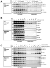
Time- and concentration-dependent DbpA secretion following LPS and ionomycin stimulation of rat mesangial and A375 melanoma cells. (A) An LPS concentration-dependent DbpA secretion takes place in rMCs. While DbpA_b is constantly secreted, DbpA_a appears around 4 h and then disappears after 12 h. (B) DbpA is secreted in a concentration-dependent manner by rMCs with a maximum at 100 ng/mL LPS for 8 h. Melanoma A375 cells do not secrete DbpA in response to LPS stimulation. (C) Both rMCs and A375 show a concentration-dependent secretion of DbpA_a/b following ionomycin stimulation for 4 h. (D) DbpA secretion in rat mesangial cells (rMCs) following LPS incubation is enhanced after preincubation with Brefeldin A, an inhibitor of the classical secretory pathway. In melanoma A375 cells, brefeldin A preincubation before ionomycin stimulation has no effect on DbpA secretion. (E) Monensin, another inhibitor of the classical secretory pathway, induces DbpA secretion in rMCs and enhances the amount of secreted DbpA when combined with LPS. DbpA secretion in response to ionomycin stimulation is not reduced in melanoma A375 cells preincubated with monensin. Each experiment was performed at least 2 times. The asterisks indicate bands from the previous antibody. DbpA, DNA-binding protein A; LPS, lipopolysaccharide; rMCs, rat mesangial cells; sc, solvent control; YB-1, Y-box-binding protein 1.
Figure 2
Figure 2
Time- and concentration-dependent DbpA secretion following LPS and ionomycin stimulation of rat mesangial and A375 melanoma cells. (A) An LPS concentration-dependent DbpA secretion takes place in rMCs. While DbpA_b is constantly secreted, DbpA_a appears around 4 h and then disappears after 12 h. (B) DbpA is secreted in a concentration-dependent manner by rMCs with a maximum at 100 ng/mL LPS for 8 h. Melanoma A375 cells do not secrete DbpA in response to LPS stimulation. (C) Both rMCs and A375 show a concentration-dependent secretion of DbpA_a/b following ionomycin stimulation for 4 h. (D) DbpA secretion in rat mesangial cells (rMCs) following LPS incubation is enhanced after preincubation with Brefeldin A, an inhibitor of the classical secretory pathway. In melanoma A375 cells, brefeldin A preincubation before ionomycin stimulation has no effect on DbpA secretion. (E) Monensin, another inhibitor of the classical secretory pathway, induces DbpA secretion in rMCs and enhances the amount of secreted DbpA when combined with LPS. DbpA secretion in response to ionomycin stimulation is not reduced in melanoma A375 cells preincubated with monensin. Each experiment was performed at least 2 times. The asterisks indicate bands from the previous antibody. DbpA, DNA-binding protein A; LPS, lipopolysaccharide; rMCs, rat mesangial cells; sc, solvent control; YB-1, Y-box-binding protein 1.
Figure 3
Figure 3
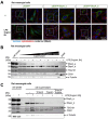
DbpA secretion requires inflammasome activation. (A) rMCs and melanoma A375 cells were either left untreated or were pretreated with inhibitor EGTA (1.5 mM) or solvent (sc), as indicated, before stimulating the cells with ionomycin for 4 h. Cell supernatants were collected, acetone precipitated, and then separated by gel electrophoresis. The presence of extracellular DbpA or YB-1 is determined by immunoblotting using the antibodies indicated. Recombinant proteins were included to determine the position of the proteins (see Supplementary Figures S3 and S4). Each experiment was performed at least 2 times. *, Nonspecific band. (B) MCC950 (1 µM). (C) Glyburide (5 µM). (D) Probenecid (2 µM). (E) Reserpine (20 µg/µL). DbpA, DNA-binding protein A; LPS, lipopolysaccharide; rMCs, rat mesangial cells; sc, solvent control; YB-1, Y-box-binding protein 1.
Figure 4
Figure 4
Cold shock proteins are secreted independently of one another. Murine bone-marrow-derived macrophages (BMDMs) from wild-type (Ybx3+/+) and Ybx3-deficient (Ybx3−/−) mice were kept in culture or challenged with the indicated stimuli. Cell supernatants were precipitated with acetone, separated on polyacrylamide gels, and transferred onto membranes. Cold shock protein content was determined by probing with the indicated antibodies. The cytoskeletal protein α-tubulin was included to control for cell lysis.
Figure 5
Figure 5
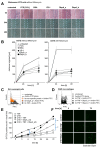
DbpA is released as a non-vesicular protein. (A) Fluorescence microscopy revealed GFP-DbpA_a and DbpA_b enriched vesicles in rMCs, transfected with both GFP-DbpA_a and DbpA_b, followed by LPS stimulation for 6 h. No vesicles were detected in GFP-transfected control cells. Scale bar is 50 µm. (B) Isolation of the exosomal fraction from LPS-treated rMC supernatants by differential ultracentrifugation showed that no DbpA_a and only a tiny amount of DbpA_b was found in the exosomes, suggesting that DbpA secretion does not involve membrane vesicles. (C) Supernatants of LPS-treated rMCs were incubated with the detergent Triton X100, which may disrupt lipid vesicles, and/or the protease trypsin. Trypsin alone digested DbpA, showing that DbpA is not protected by vesicular structures and is released as a non-vesicular protein. Thus, we conclude that DbpA is not present in exosomes. Each experiment was performed at least 2 times. DbpA, DNA-binding protein A; GFP, green fluorescent protein; LPS, lipopolysaccharide; rMCs, rat mesangial cells; TSG101, tumor susceptibility gene 101.
Figure 6
Figure 6
Extracellular DbpA promotes cell migration, binds to cell membranes, and delays cytotoxic TNF effects. (A) The wound closing capacity of melanoma A375 cells, incubated with and without mitomycin, an inhibitor of cell proliferation, shows enhanced wound closure following application of recombinant cold shock proteins (YB-1, DbpA_a, and DbpA_b (each 1 µg/mL)). BSA (1 µg/mL, negative control), FCS (10%, positive control). (B) Quantification of cell migration in response to the stimuli shown in (A) is presented. (C) Biotinylated recombinant human DbpA demonstrates cell surface binding on rMCs. An unspecific biotinylated protein was included as the negative control and biotinylated TNF as the positive control. (D) Recombinant human DbpA interferes with the binding of biotinylated TNF to RAW macrophages. Avidin–FITC alone was included as the negative control and biotinylated TNF as the positive control. (E,G) DbpA modifies the death-inducing activity of TNF. HK-2 cells (E) or mouse PTECs (G) were either left untreated or incubated overnight with cycloheximide (CHX). Cells were then washed, SYTOX green was applied, and the cells were treated as indicated. Cell death was determined by the number of SYTOX positive nuclei appearing over time. (F,H) Data were analyzed using one-way ANOVA. * p < 0.05. Scale bar is 50 µm. Each experiment was performed at least 3 times. BSA, bovine serum albumin; CHX, cycloheximide; DbpA, DNA-binding protein A; HK-2, human kidney 2; TNF, tumor necrosis factor; PTEC, primary tubular epithelial cell; YB-1, Y-box-binding protein 1.
Figure 6
Figure 6
Extracellular DbpA promotes cell migration, binds to cell membranes, and delays cytotoxic TNF effects. (A) The wound closing capacity of melanoma A375 cells, incubated with and without mitomycin, an inhibitor of cell proliferation, shows enhanced wound closure following application of recombinant cold shock proteins (YB-1, DbpA_a, and DbpA_b (each 1 µg/mL)). BSA (1 µg/mL, negative control), FCS (10%, positive control). (B) Quantification of cell migration in response to the stimuli shown in (A) is presented. (C) Biotinylated recombinant human DbpA demonstrates cell surface binding on rMCs. An unspecific biotinylated protein was included as the negative control and biotinylated TNF as the positive control. (D) Recombinant human DbpA interferes with the binding of biotinylated TNF to RAW macrophages. Avidin–FITC alone was included as the negative control and biotinylated TNF as the positive control. (E,G) DbpA modifies the death-inducing activity of TNF. HK-2 cells (E) or mouse PTECs (G) were either left untreated or incubated overnight with cycloheximide (CHX). Cells were then washed, SYTOX green was applied, and the cells were treated as indicated. Cell death was determined by the number of SYTOX positive nuclei appearing over time. (F,H) Data were analyzed using one-way ANOVA. * p < 0.05. Scale bar is 50 µm. Each experiment was performed at least 3 times. BSA, bovine serum albumin; CHX, cycloheximide; DbpA, DNA-binding protein A; HK-2, human kidney 2; TNF, tumor necrosis factor; PTEC, primary tubular epithelial cell; YB-1, Y-box-binding protein 1.
Figure 7
Figure 7
Scheme depicting the mechanism of ionomycin-induced cold shock protein (CSP) secretion. Ionomycin triggers intracellular calcium release that activates the inflammasome, which in turn, facilitates the secretion of intracellular cold shock proteins. The inhibition of either calcium influx or inflammasome activation blocks stimulus-induced protein secretion. The analysis of Ybx3-deficient cells shows that cold shock protein secretion occurs independent of one another. Taken together, our data suggest that both proteins aggregate in the serum to form a complex, which is also found in the urine following kidney injury. Since cold shock proteins bind RNA and DNA, we hypothesize that extracellular RNA/DNA may act as a stabilizing factor that explains the observed stoichiometry of 10 DbpA per 1 YB-1 molecule.

References

    1. Espinosa-Cantu A., Cruz-Bonilla E., Noda-Garcia L., DeLuna A. Multiple forms of multifunctional proteins in health and disease. Front. Cell Dev. Biol. 2020;8:451. doi: 10.3389/fcell.2020.00451. - DOI - PMC - PubMed
    1. Chen R., Niu L., Wu L., He Y., Liu G., Hong K. Identification of an endoplasmic reticulum stress-associated gene signature to predict the immune status and prognosis of cutaneous melanoma. Medicine. 2022;101:e30280. doi: 10.1097/MD.0000000000030280. - DOI - PMC - PubMed
    1. Lindquist J.A., Mertens P.R. Cold shock proteins: From cellular mechanisms to pathophysiology and disease. Cell Commun. Signal. 2018;16:63. doi: 10.1186/s12964-018-0274-6. - DOI - PMC - PubMed
    1. Kohno K., Izumi H., Uchiumi T., Ashizuka M., Kuwano M. The pleiotropic functions of the y-box-binding protein, yb-1. Bioessays. 2003;25:691–698. doi: 10.1002/bies.10300. - DOI - PubMed
    1. Wolffe A.P. Structural and functional properties of the evolutionarily ancient y-box family of nucleic acid binding proteins. Bioessays. 1994;16:245–251. doi: 10.1002/bies.950160407. - DOI - PubMed

Publication types

MeSH terms

LinkOut - more resources