Comparative Study
. 2024 Oct 21;13(20):1745.
doi: 10.3390/cells13201745.

Comparative Analysis of Extracellular Vesicles from Cytotoxic CD8+ αβ T Cells and γδ T Cells

Affiliations
Comparative Study

Comparative Analysis of Extracellular Vesicles from Cytotoxic CD8+ αβ T Cells and γδ T Cells

Lisa Griesel et al. Cells. .

Abstract

Background: Although belonging to different branches of the immune system, cytotoxic CD8+ αβ T cells and γδ T cells utilize common cytolytic effectors including FasL, granzymes, perforin and granulysin. The effector proteins are stored in different subsets of lysosome-related effector vesicles (LREVs) and released to the immunological synapse upon target cell encounter. Notably, in activated cells, LREVs and potentially other vesicles are continuously produced and released as extracellular vesicles (EVs). Presumably, EVs serve as mediators of intercellular communication in the local microenvironment or at distant sites.

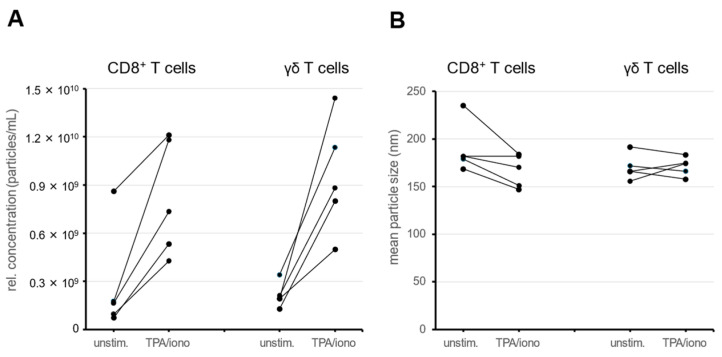
Methods: EVs of activated and expanded cytotoxic CD8+ αβ T cells or γδ T cells were enriched from culture supernatants by differential and ultracentrifugation and characterized by nanoparticle tracking analyses and Western blotting. For a comparative proteomic profiling, EV preparations from both cell types were isobaric labeled with tandem mass tags (TMT10plex) and subjected to mass spectrometry analysis.

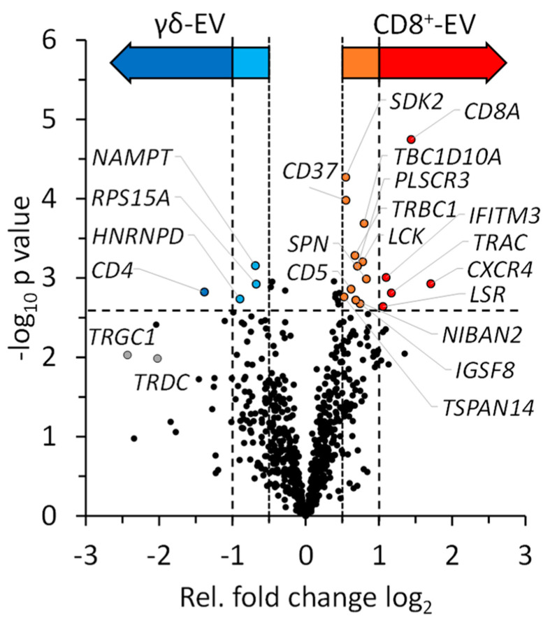
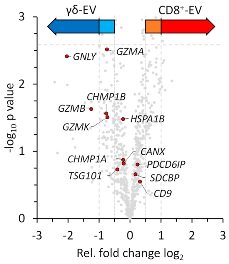
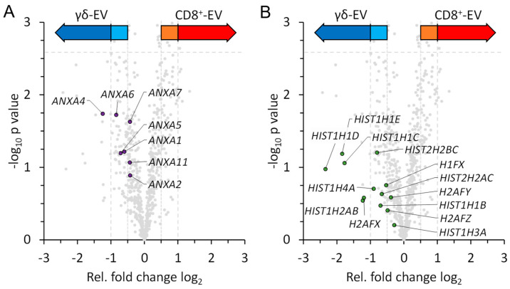
Results: 686 proteins were quantified in EV preparations of cytotoxic CD8+ αβ T cells and γδ T cells. Both populations shared a major set of similarly abundant proteins, while much fewer proteins presented higher abundance levels in either CD8+ αβ T cells or γδ T cells. To our knowledge, we provide the first comparative analysis of EVs from cytotoxic CD8+ αβ T cells and γδ T cells.

Keywords: CD8+ T cells; cytotoxic T cells; cytotoxic effector proteins; cytotoxic granules; exosomes; extracellular vesicles (EVs); lysosome-related effector vesicles (LREVs); proteomics profiling; αβ T cells; γδ T cells.


Conflict of interest statement

The authors declare no conflicts of interest.

Figures

Figure 1
Figure 1
CD8+ αβ T cells or γδ T cells were left unstimulated (unstim.) or stimulated for 2 h with phorbol ester and ionomycin (TPA/iono), before EVs were isolated by differential centrifugation and ultracentrifugation and the particle concentration (A) and particle size (B) were analyzed by NTA. Data for five individual experiments with CD8+ αβ T cells or γδ T cells are shown. If less than 4 × 10⁸ cells were plated, the particle concentration was extrapolated respectively for unstimulated and stimulated cells.
Figure 2
Figure 2
PHA-expanded CD8+ αβ T cells (squares and thin lines) and zoledronate-expanded γδ T cells (circles and thick lines) were left untreated or exposed to TPA and ionomycin for six hours. Every hour, cell viability was determined by flow cytometry using far red stain (FRS) exclusion.
Figure 3
Figure 3
(A) Mean particle size. (B) Relative particle concentration. CD8+ and γδ T cell-derived EVs were subjected to NTA with four individual track measurements for each sample. The cells were stimulated or not with TPA and ionomycin for 2 h (n = 5 for CD8+ and n = 5 for γδ+ T cells). Alternatively, cells were left untreated for 72 h (n = 8 for CD8+ and n = 7 for γδ+ T cells). If less than 4 × 10⁸ cells were plated, the particle concentration was extrapolated accordingly.
Figure 4
Figure 4
Quantitative analysis of EVs from expanded CD8+ T cells and zoledronate-stimulated γδ T cells. Individual proteins with significant fold-change differences are marked and indicated by their gene names within the volcano plot. In addition, we highlighted the TCR γ and δ chains although their fold-change difference did not reach significance. The color code of the arrows corresponds to Supplementary Table S2: Dark Red – Significantly higher abundant in EVs of CD8+ T cells (Strict); Light Red—Significantly higher abundant in EVs of CD8+ T cells (Moderate); Dark Blue—Significantly higher abundant in EVs of γδ T cells (Strict); Light Blue—Significantly higher abundant in EVs of γδ T cells (Moderate).
Figure 5
Figure 5
Quantitative analysis of EVs from expanded CD8+ T cells and zoledronate-stimulated γδ T cells. Individual EV-associated markers and effector proteins are highlighted and indicated by their gene names within the volcano plot. Color code as described in Figure 4.
Figure 6
Figure 6
Quantitative analysis of EVs from expanded CD8+ T cells and zoledronate-stimulated γδ T cells. Individual annexins (A) and histones (B), which were more abundant in EVs from γδ T cells, are highlighted and indicated by their gene names within the volcano plot. Color code as described in Figure 4.

Similar articles

References

    1. Lettau M., Kabelitz D., Janssen O. Lysosome-Related Effector Vesicles in T Lymphocytes and NK Cells. Scand. J. Immunol. 2015;82:235–243. doi: 10.1111/sji.12337. - DOI - PubMed
    1. Blott E.J., Griffiths G.M. Secretory lysosomes. Nat. Rev. Mol. Cell Biol. 2002;3:122–131. doi: 10.1038/nrm732. - DOI - PubMed
    1. Peters P.J., Borst J., Oorschot V., Fukuda M., Krähenbühl O., Tschopp J., Slot J.W., Geuze H.J. Cytotoxic T lymphocyte granules are secretory lysosomes, containing both perforin and granzymes. J. Exp. Med. 1991;173:1099–1109. doi: 10.1084/jem.173.5.1099. - DOI - PMC - PubMed
    1. Blott E.J., Bossi G., Clark R., Zvelebil M., Griffiths G.M. Fas ligand is targeted to secretory lysosomes via a proline-rich domain in its cytoplasmic tail. J. Cell Sci. 2001;114:2405–2416. doi: 10.1242/jcs.114.13.2405. - DOI - PubMed
    1. Lettau M., Schmidt H., Kabelitz D., Janssen O. Secretory lysosomes and their cargo in T and NK cells. Immunol. Lett. 2007;108:10–19. doi: 10.1016/j.imlet.2006.10.001. - DOI - PubMed

Publication types

MeSH terms

Substances

LinkOut - more resources