Review
. 2024 Sep 24;11(10):954.
doi: 10.3390/bioengineering11100954.

Bridging the Gap: Advances and Challenges in Heart Regeneration from In Vitro to In Vivo Applications

Affiliations
Review

Bridging the Gap: Advances and Challenges in Heart Regeneration from In Vitro to In Vivo Applications

Tatsuya Watanabe et al. Bioengineering (Basel). .

Abstract

Cardiovascular diseases, particularly ischemic heart disease, area leading cause of morbidity and mortality worldwide. Myocardial infarction (MI) results in extensive cardiomyocyte loss, inflammation, extracellular matrix (ECM) degradation, fibrosis, and ultimately, adverse ventricular remodeling associated with impaired heart function. While heart transplantation is the only definitive treatment for end-stage heart failure, donor organ scarcity necessitates the development of alternative therapies. In such cases, methods to promote endogenous tissue regeneration by stimulating growth factor secretion and vascular formation alone are insufficient. Techniques for the creation and transplantation of viable tissues are therefore highly sought after. Approaches to cardiac regeneration range from stem cell injections to epicardial patches and interposition grafts. While numerous preclinical trials have demonstrated the positive effects of tissue transplantation on vasculogenesis and functional recovery, long-term graft survival in large animal models is rare. Adequate vascularization is essential for the survival of transplanted tissues, yet pre-formed microvasculature often fails to achieve sufficient engraftment. Recent studies report success in enhancing cell survival rates in vitro via tissue perfusion. However, the transition of these techniques to in vivo models remains challenging, especially in large animals. This review aims to highlight the evolution of cardiac patch and stem cell therapies for the treatment of cardiovascular disease, identify discrepancies between in vitro and in vivo studies, and discuss critical factors for establishing effective myocardial tissue regeneration in vivo.

Keywords: 3D printing; cardiac regeneration; heart tissue; microvasculature; paracrine effect; transplantation; vascularization.


Conflict of interest statement

The authors declare no conflicts of interest.

Figures

Figure 1
Figure 1
Perfusable vascular networks in 3D engineered tissue. (A) An approach by multi-scale vascular networks (MSVTs) comprised of patterned macro-channels integrated with self-assembled micro-vessels, within 3D engineered tissues. The engineered macro-vessels anastomose with the surrounding micro-vasculature can be observed in histological images (white arrows indicate micro-vessels sprouts; dashed lines indicate the macro-vessels borders). (white bar: 100 μm (left), 50 μm (right)). (B) Millimetric vessel-like scaffolds and 3D bioprinted vascularized tissues interconnect, creating fully engineered hierarchical vascular constructs for implantation. The spontaneously formed vessels in the hydrogel communicate with the endothelium through the scaffold fenestrations, enabling the microvasculature perfusion. Representative confocal images of assembled vascular constructs with endothelium ECs (dTomato-ECs, red) and printed microvascular ECs (ZsGreen-ECs, green) after one week in culture (white bar: 100 μm). (C) Sacrificial writing into functional tissue (SWIFT). Perfusable EB tissue fabricated by SWIFT. An image sequence showing the embedded 3D printing of a branched, hierarchical vascular network within a compacted EB-based tissue matrix connected to inlet and outlet tubes, seen entering the tissue from the left and right (white bar: 10 mm). Image of the perfusable tissue construct after 12 h of perfusion and fluorescent image of LIVE/DEAD (green/red) cell viability stains at various sections through the tissue (white bar: 10 mm). Reprinted from refs. [40,63,95] with Copyright permission.
Figure 2
Figure 2
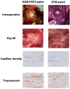
Effects of Growth Factor-Enhanced Patch. Compared to the ECM patch, the ECM patch supplemented with FGF2 (Fibroblast Growth Factor 2) has shown an increase in capillary density and a higher number of tropomyosin-positive cells. Adapted from ref. [14].
Figure 3
Figure 3
Differences in vascular formation and fibrosis after cardiac patch transplantation in thymectomized mice and rats. In thymectomized mice, a well-established vascular structure with mouse blood components (TER119) within the vessels is observed (White arrows indicate vascular cord). Additionally, Sirius red staining indicates relatively mild fibrosis in the transplanted cardiac patch. Conversely, similar experiments in thymectomized rats show poor vascular development and significant fibrosis in the transplanted patch. Adapted from ref. [115].

Similar articles

Cited by

References

    1. Anderson J.L., Morrow D.A. Acute Myocardial Infarction. N. Engl. J. Med. 2017;376:2053–2064. doi: 10.1056/NEJMra1606915. - DOI - PubMed
    1. Lund L.H., Edwards L.B., Kucheryavaya A.Y., Benden C., Christie J.D., Dipchand A.I., Dobbels F., Goldfarb S.B., Levvey B.J., Meiser B., et al. The registry of the International Society for Heart and Lung Transplantation: Thirty-first official adult heart transplant report—2014; focus theme: Retransplantation. J. Heart Lung Transplant. 2014;33:996–1008. doi: 10.1016/j.healun.2014.08.003. - DOI - PubMed
    1. Mozaffarian D., Benjamin E.J., Go A.S., Arnett D.K., Blaha M.J., Cushman M., Das S.R., de Ferranti S., Despres J.-P., Fullerton H.J., et al. Heart Disease and Stroke Statistics-2016 Update: A Report From the American Heart Association. Circulation. 2016;133:e38–e360. doi: 10.1161/CIR.0000000000000350. - DOI - PubMed
    1. Porrello E.R., Mahmoud A.I., Simpson E., Hill J.A., Richardson J.A., Olson E.N., Sadek H.A. Transient regenerative potential of the neonatal mouse heart. Science. 2011;331:1078–1080. doi: 10.1126/science.1200708. - DOI - PMC - PubMed
    1. Bergmann O., Bhardwaj R.D., Bernard S., Zdunek S., Barnabé-Heider F., Walsh S., Zupicich J., Alkass K., Buchholz B.A., Druid H., et al. Evidence for cardiomyocyte renewal in humans. Science. 2009;324:98–102. doi: 10.1126/science.1164680. - DOI - PMC - PubMed

LinkOut - more resources