

The Buccal Fat Pad: A Unique Human Anatomical Structure and Rich and Easily Accessible Source of Mesenchymal Stem Cells for Tissue Repair
- PMID: 39451344
- PMCID: PMC11505344
- DOI: 10.3390/bioengineering11100968
The Buccal Fat Pad: A Unique Human Anatomical Structure and Rich and Easily Accessible Source of Mesenchymal Stem Cells for Tissue Repair
Abstract
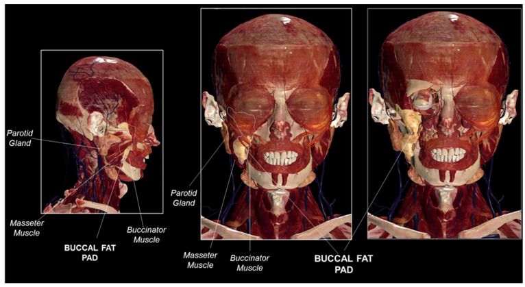
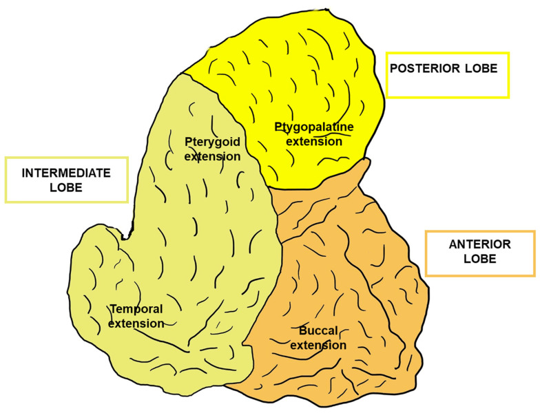
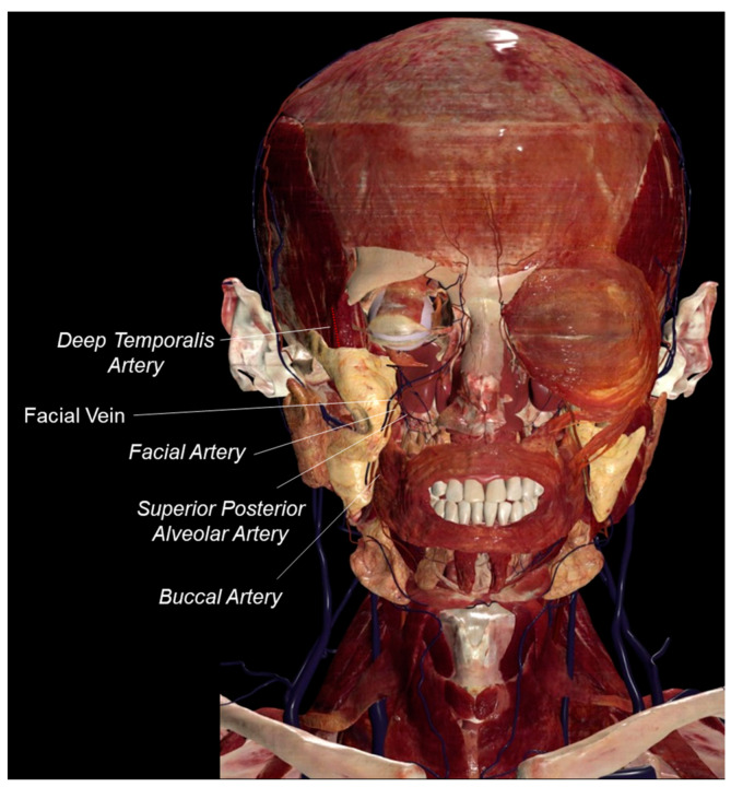
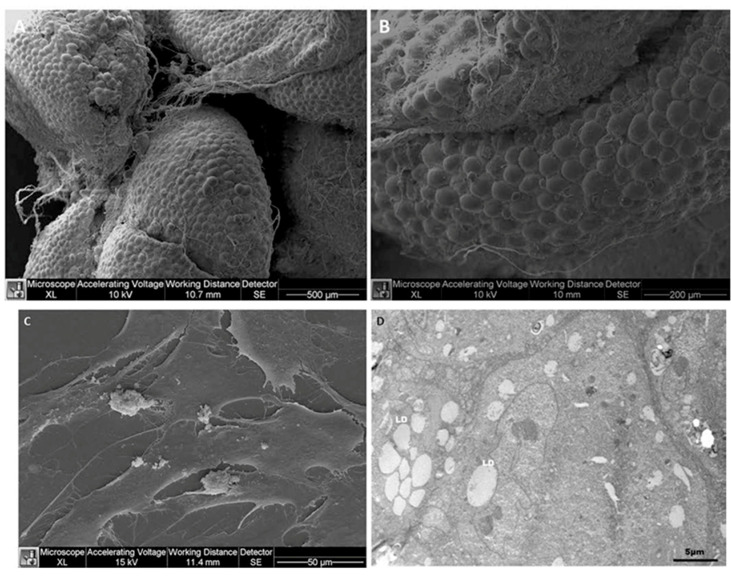
Buccal fat pads are biconvex adipose tissue bags that are uniquely found on both sides of the human face along the anterior border of the masseter muscles. Buccal fat pads are important determinants of facial appearance, facilitating gliding movements of facial masticatory and mimetic muscles. Buccal fad pad flaps are used for the repair of oral defects and as a rich and easily accessible source of mesenchymal stem cells. Here, we introduce the buccal fat pad anatomy and morphology and report its functions and applications for oral reconstructive surgery and for harvesting mesenchymal stem cells for clinical use. Future frontiers of buccal fat pad research are discussed. It is concluded that many biological and molecular aspects still need to be elucidated for the optimal application of buccal fat pad tissue in regenerative medicine.
Keywords: buccal fat pad; human anatomy; mesenchymal stem cells source; oral surgery.
Conflict of interest statement
The authors declare no conflicts of interest.
Figures
Similar articles
-
Anatomical structure of the buccal fat pad and its clinical adaptations.Plast Reconstr Surg. 2002 Jun;109(7):2509-18; discussion 2519-20. doi: 10.1097/00006534-200206000-00052. Plast Reconstr Surg. 2002. PMID: 12045584
-
Buccal Fat Pad Reduction.2022 Nov 21. In: StatPearls [Internet]. Treasure Island (FL): StatPearls Publishing; 2025 Jan–. 2022 Nov 21. In: StatPearls [Internet]. Treasure Island (FL): StatPearls Publishing; 2025 Jan–. PMID: 35015438 Free Books & Documents.
-
Porcine adipose-derived stem cells from buccal fat pad and subcutaneous adipose tissue for future preclinical studies in oral surgery.Stem Cell Res Ther. 2013;4(6):148. doi: 10.1186/scrt359. Stem Cell Res Ther. 2013. PMID: 24330736 Free PMC article.
-
The buccal pad of fat: a review.Clin Anat. 1995;8(6):403-6. doi: 10.1002/ca.980080606. Clin Anat. 1995. PMID: 8713160 Review.
-
Anatomy of the Buccal Space: Surgical and Radiological Perspectives.J Craniofac Surg. 2024 Oct 1;35(7):1972-1976. doi: 10.1097/SCS.0000000000010411. Epub 2024 Jun 17. J Craniofac Surg. 2024. PMID: 38885157 Review.
References
-
- Bichat X. Anatomie Générale, Applique’e la Physiologie et Àla Médecine. Brosson; Paris, France: 1801.
Publication types
Grants and funding
LinkOut - more resources
Full Text Sources
