Review
. 2024 Sep 29;11(10):983.
doi: 10.3390/bioengineering11100983.

Manufacturing, Processing, and Characterization of Self-Expanding Metallic Stents: A Comprehensive Review

Affiliations
Review

Manufacturing, Processing, and Characterization of Self-Expanding Metallic Stents: A Comprehensive Review

Saeedeh Vanaei et al. Bioengineering (Basel). .

Abstract

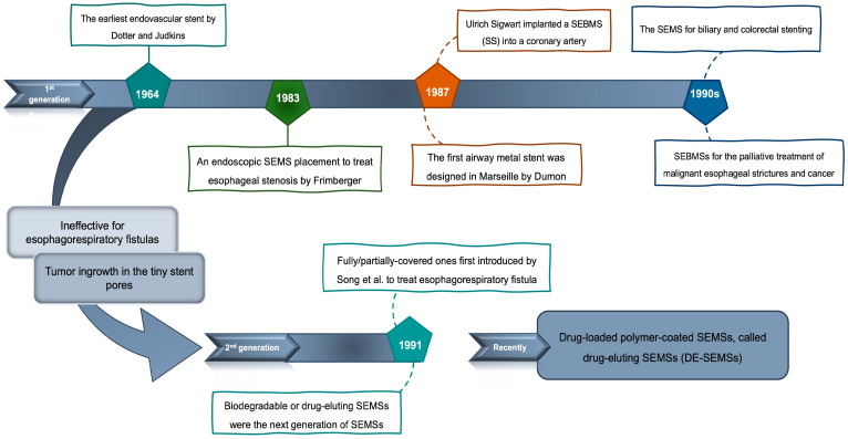
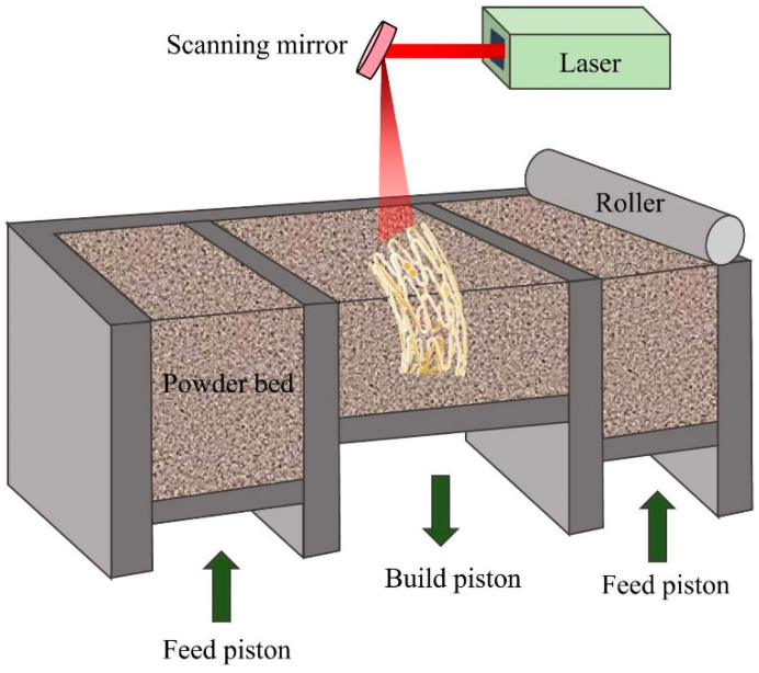
This paper aims to review the State of the Art in metal self-expanding stents made from nitinol (NiTi), showing shape memory and superelastic behaviors, to identify the challenges and the opportunities for improving patient outcomes. A significant contribution of this paper is its extensive coverage of multidisciplinary aspects, including design, simulation, materials development, manufacturing, bio/hemocompatibility, biomechanics, biomimicry, patency, and testing methodologies. Additionally, the paper offers in-depth insights into the latest practices and emerging trends, with a special emphasis on the transformative potential of additive manufacturing techniques in the development of metal stents. By consolidating existing knowledge and highlighting areas for future innovation, this review provides a valuable roadmap for advancing nitinol stents.

Keywords: additive manufacturing; biomaterials; biomimicry; patency rate; self-expanding metallic stents; shape memory alloy.


Conflict of interest statement

The authors declare no conflict of interest.

Figures

Figure 1
Figure 1
A timeline for the development of the stents from early 1960s. The evolution from the first to the second generation is depicted.
Figure 2
Figure 2
(a) Mg alloy stent. Reproduced from open access journals [40] (b-I) braided NiTi stent, (b-II) laser cut NiTi stent. Reprinted with permission [41]. Copyright 2021, Elsevier. (c) Cobalt alloy (d) Tantalum alloy stent. Reprinted with permission ([42]). Copyright 2009, Taylor & Francis.
Figure 3
Figure 3
Classifications of metallic stents based on different terms.
Figure 4
Figure 4
Classifications of coatings used for metal stents.
Figure 5
Figure 5
Schematic illustration of hot extrusion and cold tube drawing processes.
Figure 6
Figure 6
A schematic illustration of the electroforming method for manufacturing pure iron stent. Reproduced with permission [30]. Copyright 2022, Elsevier.
Figure 7
Figure 7
Schematic illustrations of textile construction methods for manufacturing metal stents: (a) braiding and (b) knitting. Reproduced with permission [30]. copyright 2019, Elsevier.
Figure 8
Figure 8
Manufacturing methods of SEMSs.
Figure 9
Figure 9
Designs of stents: (a) slotted tube, (b) coiled stent, (c) braided stent, (d) knitted stent, and (e) helical. Reproduced with permission [30]. Copyright 2022, Elsevier. (f) covered, (g) uncovered stents. Reproduced with permission [95]. Copyright 2010, Elsevier.
Figure 10
Figure 10
Surface characteristics of NiTi stents: bare NiTi stent struts at (a-I) low and (a-II) high magnification. Reprinted from open access journals [106]. (b) stent manufactured by LPBF. Reprinted from open access journals [107], (c) surface after mechanical polish, (d) after passivation, and (e) after electropolishing [108]. Copyright 2006, Elsevier.
Figure 11
Figure 11
Different surface characteristics of stents. This illustration shows that biocompatibility and corrosion resistance are affected by the post-surface treatments.
Figure 12
Figure 12
Stress–strain–temperature diagram of NiTi. Reproduced with permission [136]. Copyright 2013, Elsevier.
Figure 13
Figure 13
A finite element model utilizing the COMSOL Multiphysics® numerical software has been constructed for the Palmaz Schatz stent, focusing on analyzing one twenty-fourth of the stent’s geometry. Reproduced with permission [196]. Copyright 2021, Elsevier.
Figure 14
Figure 14
Stent size variation: (a) unit cell, (b) 2D pattern of cell, and (c) stent. Reproduced with permission [197]. Copyright 2020, Elsevier.
Figure 15
Figure 15
(a) A thorough investigation into post-processing analyses has been undertaken for four specific types of expandable stents, namely PLLA, nitinol, stainless steel (SS), and pure Mg, (b) along with an assessment covering six distinct geometrical variations in these stents. (c) These analyses encompass the evaluation of area percentages via histograms, with a primary focus on instances of adverse low WSS (<0.5 Pa) at four critical time points during a cardiac cycle. (d) Contour maps illustrate the distribution of time-averaged wall shear stress (TAWSS) on the lumen wall. Reproduced with permission [199]. Copyright 2019, Frontiers.
Figure 16
Figure 16
(a) Normalized effective wall shear stress (WSS), (b) normalized average axial WSS, (c) normalized average transverse WSS, and (d) ratio of normalized axial WSS to transverse WSS plotted for the various stent design types. Additionally, the percentage area of the region between struts with averaged low WSS ((e) <5 dynes/cm2 and (f) <2.5 dynes/cm2) for more than 50% of the flow cycle in the different stent design types. Reproduced with permission [204]. Copyright 2009, The American Society of Mechanical Engineers.
Figure 17
Figure 17
(a) This section presents a comprehensive comparative analysis of radial-force responses, drawing from both experimental and computational data, for wire-braided stents with braid angles of α = 45°. (b) Juxtapose computational bending deformations. (c) Comparison of experimental and computational data. (d) Stent elongation at 2.4 mm. Reproduced with permission [205]. Copyright 2021, Elsevier.
Figure 17
Figure 17
(a) This section presents a comprehensive comparative analysis of radial-force responses, drawing from both experimental and computational data, for wire-braided stents with braid angles of α = 45°. (b) Juxtapose computational bending deformations. (c) Comparison of experimental and computational data. (d) Stent elongation at 2.4 mm. Reproduced with permission [205]. Copyright 2021, Elsevier.
Figure 18
Figure 18
The stent deployment detection system, as elaborated in the research conducted by Xu et al. reproduced with permission [213]. Copyright 2020, John Wiley & Sons, Inc. (a) The experimental setup includes a stent and an RF-based sensor. The sensor is responsible for transmitting and receiving an amplitude-modulated signal, with the received signal being influenced by the shape of the stent. (b) The study workflow begins with data collection using the RF-based sensor for four distinct classes. A novel deep learning model, named StentNet, is introduced to detect stent deployment. (c) Data are collected in four different cases: no deployment (0 cm), partial deployment (1 cm), full deployment (3 cm), and full deployment with compression in the center. (d) Visualization of the four data classes shows the reflection power intensity, where darker colors represent higher reflection power.
Figure 19
Figure 19
(a) macrofluidic flow chambers modeling the left anterior descending coronary (left), carotid (middle) and femoral (right) arteries, and (b) shear rate distributions within the models: extracted from CFD models. Reproduced with permission [243]. Copyright 2024, CellPress.
Figure 20
Figure 20
SEM images depicting platelet aggregates on a carotid stent after perfusion with blood for 1 h. (a) Aggregates are observed at the intersection of the stent meshes and (b) composed of tightly packed platelets. Reproduced with permission [243]. Copyright 2024, CellPress.
Figure 21
Figure 21
Schematic of laser powder bed fusion process to fabricate stent. Reproduced with permission [30]. Copyright 2022, Elsevier.

Similar articles

Cited by

References

    1. Emre A., Sertkaya M., Akbulut S., Erbil O., Yurttutan N., Kale İ.T., Bülbüloğlu E. Self-expandable metallic stent application for the management of upper gastrointestinal tract disease. Turk. J. Surg. 2018;34:101. doi: 10.5152/turkjsurg.2017.3740. - DOI - PMC - PubMed
    1. Dutau H., Musani A.I., Plojoux J., Laroumagne S., Astoul P. The use of self-expandable metallic stents in the airways in the adult population. Expert Rev. Respir. Med. 2014;8:179–190. doi: 10.1586/17476348.2014.880055. - DOI - PubMed
    1. Ahadi F., Azadi M., Biglari M., Bodaghi M., Khaleghian A. Evaluation of coronary stents: A review of types, materials, processing techniques, design, and problems. Heliyon. 2023;9:e13575. doi: 10.1016/j.heliyon.2023.e13575. - DOI - PMC - PubMed
    1. Cheung D.Y., Lee Y.K., Yang C.H. Status and literature review of self-expandable metallic stents for malignant colorectal obstruction. Clin. Endosc. 2014;47:65–73. doi: 10.5946/ce.2014.47.1.65. - DOI - PMC - PubMed
    1. Kim J.H., Song H.-Y., Shin J.H. Malignant gastric outlet obstructions: Treatment with self-expandable metallic stents. Gut Liver. 2010;4((Suppl. S1)):S32. doi: 10.5009/gnl.2010.4.S1.S32. - DOI - PMC - PubMed

LinkOut - more resources