

De-Epithelization of the Human Amniotic Membrane Using a System Involving Ozonated Water and Ultrasound
- PMID: 39451363
- PMCID: PMC11504975
- DOI: 10.3390/bioengineering11100987
De-Epithelization of the Human Amniotic Membrane Using a System Involving Ozonated Water and Ultrasound
Abstract
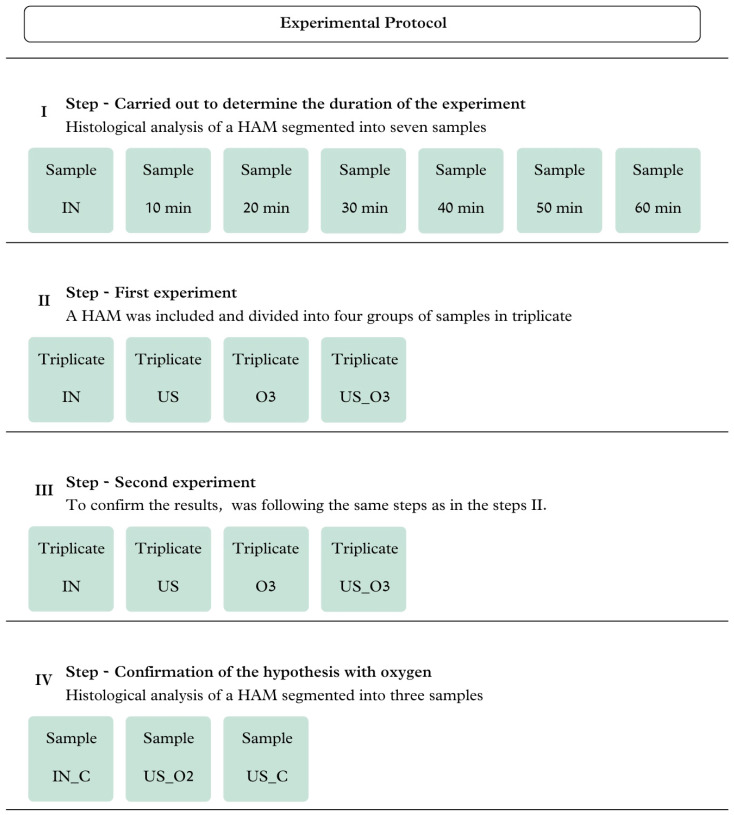
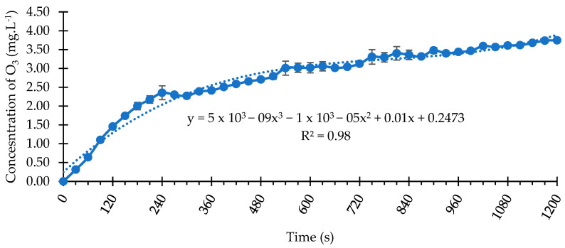
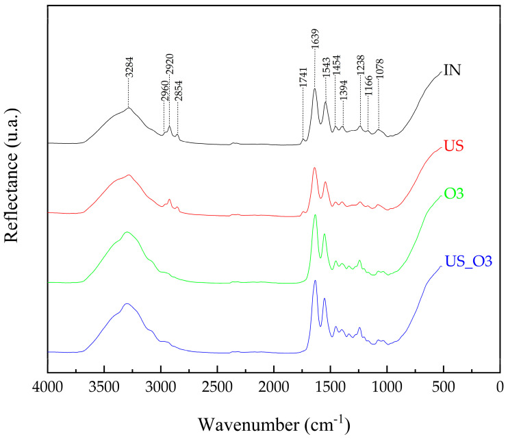
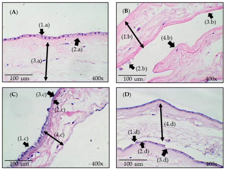
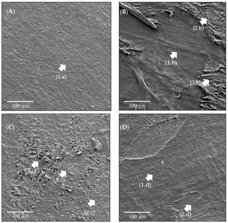
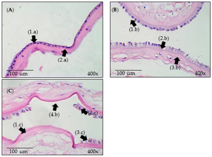
The aim of this study was to evaluate whether a system involving ozonated water and ultrasound causes de-epithelization of the human amniotic membrane (HAM). The experiment protocol was carried out in four stages. Stage I was carried out to determine the duration of the experiment. Stage II comprised the first experiment, involving four groups of samples studied in triplicate: control/natural (IN), processed with ultrasound in a liquid medium (US), processed with ozonated water (O3), and processed with ozonated water combined with ultrasound (US_O3). Stage III was performed to confirm the results, following the same steps present in Stage II. Stage IV involved the use of oxygen to confirm the hypothesis. Histological analysis was carried out to verify whether the effects of O2 were similar to those of O3. The system was activated, and ozonation was carried out for 10 min, as in the previous experiment, reaching a concentration level of 3.0 mg/L. The samples were submerged and positioned in the reservoir and processed separately for 55 min. The biochemical properties were assessed using Fourier transform infrared spectroscopy, and the morphology was examined using histology and scanning electron microscopy. The spectra of the samples exhibited similarities; however, subtle changes were highlighted, such as smooth band shifts and intensity changes. The morphology indicated that ultrasound achieved more efficient HAM de-epithelialization compared to ultrasound combined with ozonated water and ozonated water alone. One plausible hypothesis for this observation is that cavitation represents the primary mechanism responsible for de-epithelialization. When ultrasound is combined with ozone, the bubbles generated by ozone gas reduce the cavitation effect. This study is pioneering as it demonstrates an ultrasound system capable of the efficient de-epithelialization of the HAM.
Keywords: amnion; biomaterials; de-epithelization; human amniotic membrane; ozone; tissue engineering; ultrasound.
Conflict of interest statement
The authors declare no conflicts of interest.
Figures
References
-
- Munoz-Torres J.R., Martínez-González S.B., Lozano-Luján A.D., Martínez-Vázquez M.C., Velasco-Elizondo P., Garza-Veloz I., Martinez-Fierro M.L. Biological properties and surgical applications of the human amniotic membrane. Front. Bioeng. Biotechnol. 2023;10:1067480. doi: 10.3389/fbioe.2022.1067480. - DOI - PMC - PubMed
LinkOut - more resources
Full Text Sources
