. 2024 Sep 30;11(10):990.
doi: 10.3390/bioengineering11100990.

Adaptive vs. Conventional Deep Brain Stimulation: One-Year Subthalamic Recordings and Clinical Monitoring in a Patient with Parkinson's Disease

Affiliations

Adaptive vs. Conventional Deep Brain Stimulation: One-Year Subthalamic Recordings and Clinical Monitoring in a Patient with Parkinson's Disease

Laura Caffi et al. Bioengineering (Basel). .

Abstract

Conventional DBS (cDBS) for Parkinson's disease uses constant, predefined stimulation parameters, while the currently available adaptive DBS (aDBS) provides the possibility of adjusting current amplitude with respect to subthalamic activity in the beta band (13-30 Hz). This preliminary study on one patient aims to describe how these two stimulation modes affect basal ganglia dynamics and, thus, behavior in the long term. We collected clinical data (UPDRS-III and -IV) and subthalamic recordings of one patient with Parkinson's disease treated for one year with aDBS, alternated with short intervals of cDBS. Moreover, after nine months, the patient discontinued all dopaminergic drugs while keeping aDBS. Clinical benefits of aDBS were superior to those of cDBS, both with and without medications. This improvement was paralleled by larger daily fluctuations of subthalamic beta activity. Moreover, with aDBS, subthalamic beta activity decreased during asleep with respect to awake hours, while it remained stable in cDBS. These preliminary data suggest that aDBS might be more effective than cDBS in preserving the functional role of daily beta fluctuations, thus leading to superior clinical benefit. Our results open new perspectives for a restorative brain network effect of aDBS as a more physiological, bidirectional, brain-computer interface.

Keywords: adaptive deep brain stimulation; biomarkers; local field potentials; neuromodulation; parkinson’s disease; subthalamic nucleus.


Conflict of interest statement

V.A. and M.A. are employees of Newronika S.p.A. L.R., S.M., and A.P. are founders and shareholders of Newronika S.p.A. I.U.I. is a Newronika S.p.A. consultant and shareholder and received funding for research activities from Newronika S.p.A. I.U.I. received lecture honoraria and research funding from Medtronic Inc. I.U.I. is an Adjunct Professor at the Department of Neurology, NYU Grossman School of Medicine. R.E. received research fundings (paid to the Institution) by Newronika S.p.A. V.L. received payments from Medtronic Inc. for sponsored presentations, and from Wise S.R.L., Newronika S.p.A., and Boston International for consultancy services. He also received support from ClearPoint for attending meetings and holds a paid leadership role on the Boston Medical Board of Boston International.

Figures
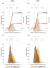
Figure 1
Figure 1
Principles of the AlphaDBS algorithm for current adjustment in adaptive mode. (a) Probability distribution of the biomarker (average normalized beta amplitude) used for current adjustment during a representative week in aDBS+ condition. Vertical dotted lines represent the biomarker limits for current adjustment (βmin and βmax). Red and black solid lines represent the stimulation current at a specific reading, respectively, for the right and left hemispheres. Current is adjusted within a predefined, clinically effective range (Amin–Amax). Numbers on top show the time percentage of the beta amplitude being less than βmin, between βmin and βmax, and above βmax in the considered week. (b) Same as (a) for the aDBS− condition. (c) Probability distribution of the biomarker during the same week in aDBS+ condition displayed in (a) separately for the awake (yellow) and asleep (brown) time. Time percentages are shown for both awake (yellow) and asleep (brown) time. (d) Same as (c) for the aDBS− condition. Abbreviations: a, adaptive; A, predefined, clinically effective amplitude; β, average normalized beta amplitude; c, conventional; DBS, deep brain stimulation; DBS+, with dopaminergic medication and DBS−, without dopaminergic medication.
Figure 2
Figure 2
Beta amplitude modulations in aDBS than in cDBS. (a) Evolution of the daily median BFRA (solid line) during awake (yellow) and asleep (brown) time. The shadowed area is bound by the daily first and third quartile of the BFRA. Vertical dotted lines represent the time points in which the treatment condition changed, as displayed in the legend on top. Grey, dark red, and purple dots mark the representative days shown, respectively, in (bd). (b) A representative day in cDBS+. Top left: daily evolution of the stimulation current for the left (black) and right (red) STN. Bottom left: daily evolution of the BFRA. Awake (8 a.m.–10 p.m.) and asleep (midnight to 6 a.m.) periods are separated by vertical solid lines. Bottom right: distribution of the BFRA separately for the awake (yellow) and asleep (brown) period. (c) Same as (b) for a representative day in aDBS+. (d) Same as (b) for a representative day in aDBS−. On these three days (bd), the total electrical energy delivered (TEED) was comparable (values reported in W. Left STN, cDBS+: 1.2 × 10⁻⁴, aDBS+: 1.3 × 10⁻⁴, aDBS−: 1.3 × 10⁻⁴; right STN, cDBS+: 1.1 × 10⁻⁴, aDBS+: 1.1 × 10⁻⁴, aDBS−: 1.1 × 10⁻⁴). In aDBS mode, the TEED was calculated every minute and then averaged across the day. (e) Boxplot of the daily median BFRA during awake (yellow) and asleep (brown) time in cDBS+, aDBS+, and aDBS−. Top horizontal lines define significant differences (solid line: p < 0.001, significance set at 0.05). (f) Same as (e) for the interquartile range of the BFRA. Abbreviations: a, adaptive; BFRA, amplitude of the STN-LFP in the patient-specific beta frequency range; c, conventional; DBS, deep brain stimulation; DBS+, with dopaminergic medication; DBS−, without dopaminergic medication; LFP, local field potential and STN, subthalamic nucleus.
Figure 3
Figure 3
Long-term stable beta-band peak frequency. (a) Median (solid line) of the daily mean amplitude spectra throughout the 11 months of recording in the three treatment conditions (cDBS+ in grey, aDBS+ in dark red, and aDBS− in purple) during awake time. The dashed area is bound by the first and third quartiles of the daily mean amplitude spectra. (b) Spectrogram of daily mean amplitude spectra during awake time. Blue vertical lines correspond to missing or removed data periods due to residual spectral artifacts after neural power law component removal (see Section 2). Black lines on top of the spectrogram represent the time course of the central frequency of the three Gaussian peaks identified in each daily mean amplitude spectrum during awake time. (c) Same as (a) during asleep time. (d) Same as (b) during asleep time. Abbreviations: a, adaptive; c, conventional; DBS, deep brain stimulation; DBS+, with dopaminergic medication and DBS−, without dopaminergic medication.

References

    1. Deuschl G., Schade-Brittinger C., Krack P., Volkmann J., Schäfer H., Bötzel K., Daniels C., Deutschländer A., Dillmann U., Eisner W., et al. A Randomized Trial of Deep-Brain Stimulation for Parkinson’s Disease. N. Engl. J. Med. 2006;355:896–908. doi: 10.1056/NEJMoa060281. - DOI - PubMed
    1. Pozzi N.G., Isaias I.U. Chapter 19—Adaptive Deep Brain Stimulation: Retuning Parkinson’s Disease. In: Quartarone A., Ghilardi M.F., Boller F., editors. Handbook of Clinical Neurology. Volume 184. Elsevier; Amsterdam, The Netherlands: 2022. pp. 273–284. Neuroplasticity. - PubMed
    1. Chen C.C., Brücke C., Kempf F., Kupsch A., Lu C.S., Lee S.T., Tisch S., Limousin P., Hariz M., Brown P. Deep Brain Stimulation of the Subthalamic Nucleus: A Two-Edged Sword. Curr. Biol. 2006;16:R952–R953. doi: 10.1016/j.cub.2006.10.013. - DOI - PubMed
    1. Rodriguez-Oroz M.C., Moro E., Krack P. Long-Term Outcomes of Surgical Therapies for Parkinson’s Disease. Mov. Disord. 2012;27:1718–1728. doi: 10.1002/mds.25214. - DOI - PubMed
    1. St George R.J., Nutt J.G., Burchiel K.J., Horak F.B. A Meta-Regression of the Long-Term Effects of Deep Brain Stimulation on Balance and Gait in PD. Neurology. 2010;75:1292–1299. doi: 10.1212/WNL.0b013e3181f61329. - DOI - PMC - PubMed

LinkOut - more resources