

Application of 3D Printing to Design and Manufacture Pancreatic Duct Stent and Animal Experiments
- PMID: 39451380
- PMCID: PMC11504459
- DOI: 10.3390/bioengineering11101004
Application of 3D Printing to Design and Manufacture Pancreatic Duct Stent and Animal Experiments
Abstract
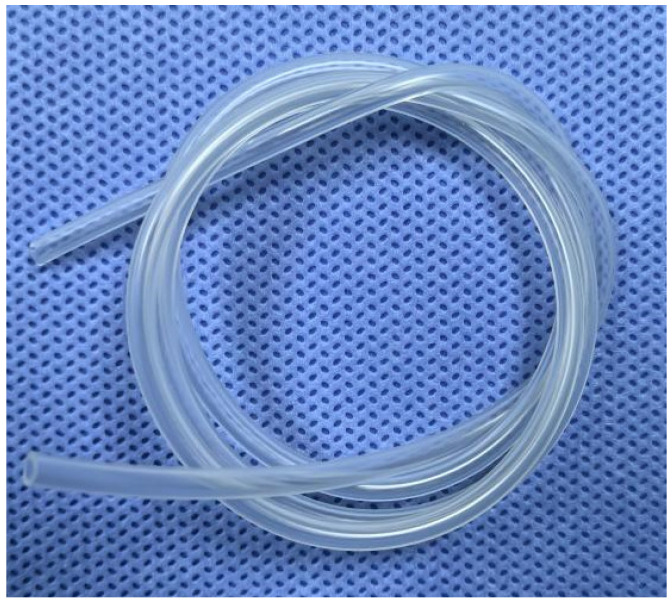
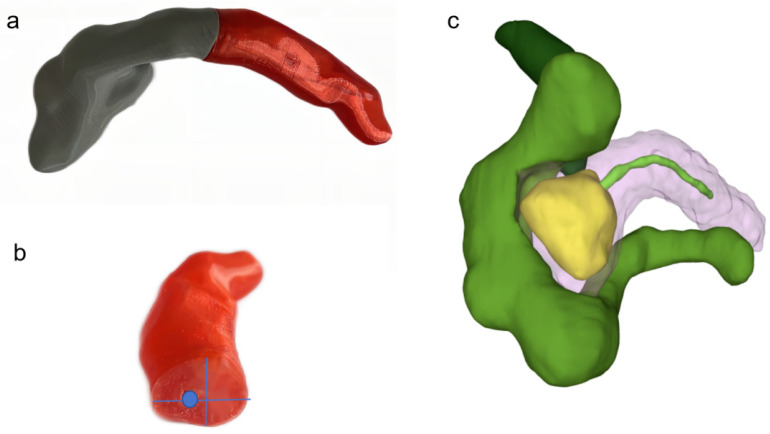
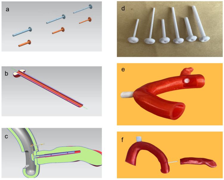
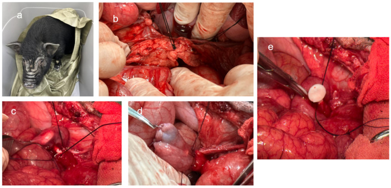
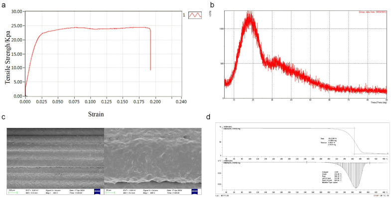
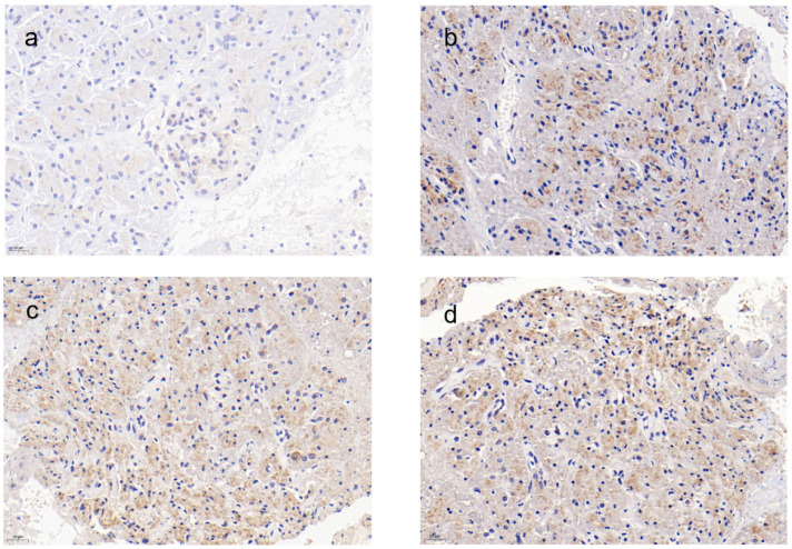
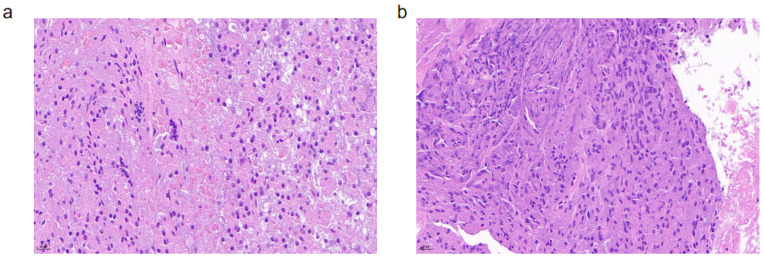
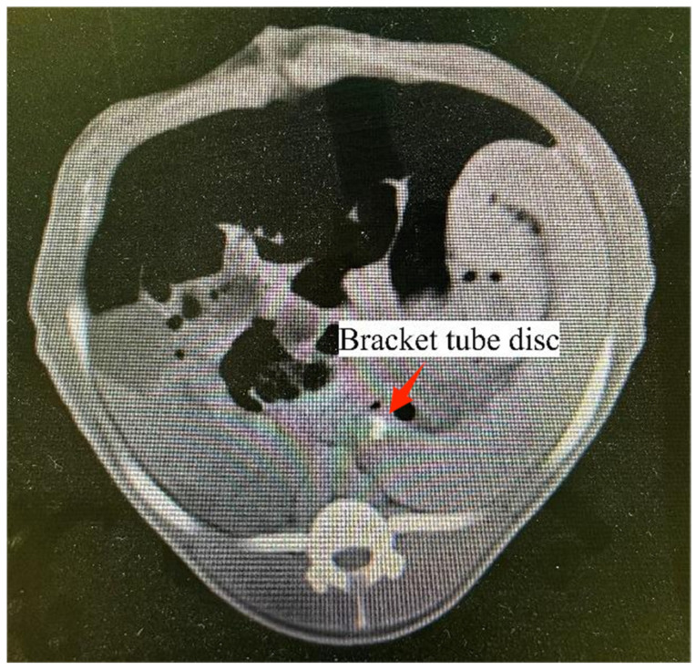
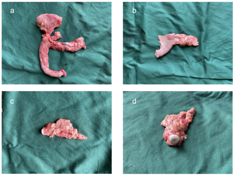
Objective: Postoperative pancreatic fistula (POPF) is a common and challenging complication following pancreaticoduodenectomy (PD), occurring in 2% to 46% of cases. Despite various pancreaticojejunostomy techniques, an effective method to prevent POPF has not been established. This study aimed to develop and evaluate a novel 3D-printed biodegradable pancreatic duct stent to simplify the surgical process of pancreaticojejunostomy, reduce anastomotic complexity, and minimize postoperative complications. Methods: Data from 32 patients undergoing total laparoscopic pancreaticoduodenectomy were utilized. Preoperative CT scans were transformed into 3D reconstructions to guide the design and printing of customized stents using polylactic acid (PLA). The stents were assessed for mechanical integrity, surface texture, and thermal stability. Animal experiments were conducted on 16 mini pigs, with the experimental group receiving the novel stent and the control group receiving traditional silicone stents. Results: The 3D-printed stents demonstrated accurate dimensional replication and mechanical reliability. In the animal experiments, the experimental group showed no significant difference in postoperative complications compared to the control group. At 4 weeks post-surgery, CT scans revealed well-healed anastomoses in both groups, with no significant inflammation or other complications. Histological examination and 3D reconstruction models confirmed good healing and device positioning in the experimental group. Conclusion: The 3D-printed biodegradable pancreatic duct stent offers a promising solution for pancreaticojejunostomy, with comparable safety and efficacy to traditional methods. Further research is needed to validate its clinical application.
Keywords: 3D printing; biodegradable stent; pancreaticoduodenectomy; pancreatojejunostomy; postoperative pancreatic fistula.
Conflict of interest statement
The authors declare no conflicts of interest.
Figures
References
-
- Pedrazzoli S. Pancreatoduodenectomy (PD) and postoperative pancreatic fistula (POPF): A systematic review and analysis of the POPF-related mortality rate in 60,739 patients retrieved from the English literature published between 1990 and 2015. Medicine. 2017;96:e6858. doi: 10.1097/MD.0000000000006858. - DOI - PMC - PubMed
-
- Kamarajah S.K., Bundred J.R., Lin A., Halle-Smith J., Pande R., Sutcliffe R., Harrison E.M., Roberts K.J., PARANOIA Study Group Systematic review and meta-analysis of factors associated with post-operative pancreatic fistula following pancreatoduodenectomy. ANZ J. Surg. 2021;91:810–821. doi: 10.1111/ans.16408. - DOI - PubMed
-
- Cao Z., Luo W., Qiu J., Liu Y., Zheng L., Zhang T. Is invagination anastomosis more effective in reducing clinically relevant pancreatic fistula for soft pancreas after pancreaticoduodenectomy under novel fistula criteria: A systematic review and meta-analysis. Front. Oncol. 2020;10:1637. doi: 10.3389/fonc.2020.01637. - DOI - PMC - PubMed
LinkOut - more resources
Full Text Sources
