. 2024 Oct 9;11(10):1005.
doi: 10.3390/bioengineering11101005.

The Use of Artificial Intelligence for Estimating Anterior Chamber Depth from Slit-Lamp Images Developed Using Anterior-Segment Optical Coherence Tomography

Affiliations

The Use of Artificial Intelligence for Estimating Anterior Chamber Depth from Slit-Lamp Images Developed Using Anterior-Segment Optical Coherence Tomography

Eisuke Shimizu et al. Bioengineering (Basel). .

Abstract

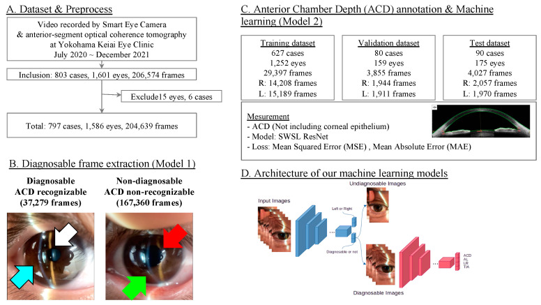
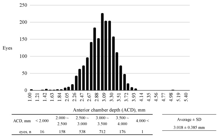
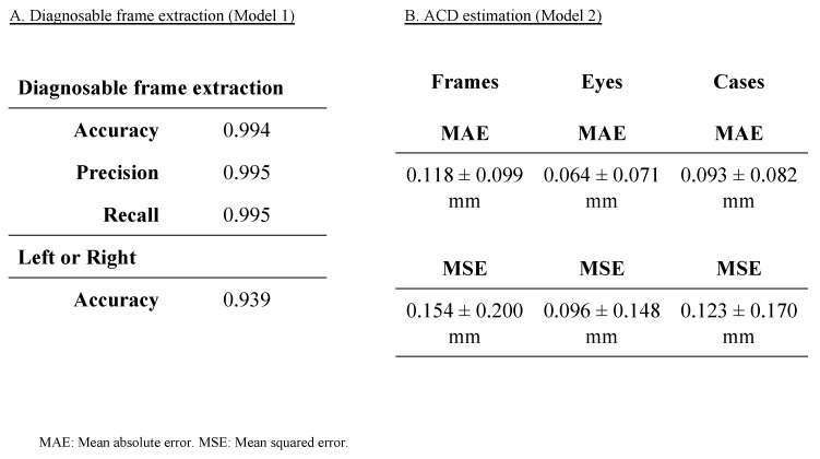
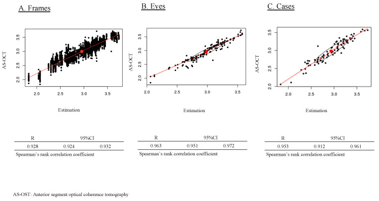
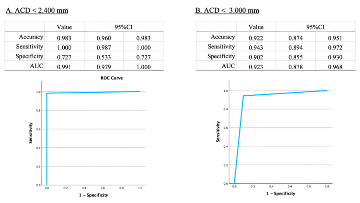
Primary angle closure glaucoma (PACG) is a major cause of visual impairment, particularly in Asia. Although effective screening tools are necessary, the current gold standard is complex and time-consuming, requiring extensive expertise. Artificial intelligence has introduced new opportunities for innovation in ophthalmic imaging. Anterior chamber depth (ACD) is a key risk factor for angle closure and has been suggested as a quick screening parameter for PACG. This study aims to develop an AI algorithm to quantitatively predict ACD from anterior segment photographs captured using a portable smartphone slit-lamp microscope. We retrospectively collected 204,639 frames from 1586 eyes, with ACD values obtained by anterior-segment OCT. We developed two models, (Model 1) diagnosable frame extraction and (Model 2) ACD estimation, using SWSL ResNet as the machine learning model. Model 1 achieved an accuracy of 0.994. Model 2 achieved an MAE of 0.093 ± 0.082 mm, an MSE of 0.123 ± 0.170 mm, and a correlation of R = 0.953. Furthermore, our model's estimation of the risk for angle closure showed a sensitivity of 0.943, specificity of 0.902, and an area under the curve (AUC) of 0.923 (95%CI: 0.878-0.968). We successfully developed a high-performance ACD estimation model, laying the groundwork for predicting other quantitative measurements relevant to PACG screening.

Keywords: Smart Eye Camera; algorithm; anterior chamber depth; anterior-segment optical coherence tomography; artificial intelligence; deep learning; glaucoma; machine learning; slit-lamp images; telemedicine.


Conflict of interest statement

OUI Inc. did not have any role in the funding, study design, data collection and analysis, decision to publish, or preparation of the manuscript. The authors declare no competing interests associated with this manuscript.

Figures

Figure 1
Figure 1
Study design and model developments. (A) Dataset and preprocessing of study chart. (B) Diagnosable frame extraction (Model 1). Images classified as diagnosable and non-diagnosable. (C) Anterior chamber depth annotation and machine learning (Model 2). Dataset is split into training, validation, and test datasets. (D) Architecture of our machine learning models.
Figure 2
Figure 2
Distribution of anterior chamber depth (ACD) in our study. The x-axis represents the ACD in millimeters (mm), ranging from 1.800 mm to 4.000 mm, divided into 0.100 mm increments. The y-axis indicates the number of eyes corresponding to each ACD measurement range. The average ACD across the study population is 3.018 ± 0.385 mm.
Figure 3
Figure 3
Performance metrics for diagnosable frame extraction (Model 1) and anterior chamber depth (ACD) estimation (Model 2). Figure 3 presents the performance metrics for the two models used in the study. Model 1 focuses on extracting diagnosable frames. Model 2 evaluates the accuracy of ACD estimation.
Figure 4
Figure 4
Correlation between AS-OCT anterior chamber depth (ACD) values and AI-estimated ACD measurements. Figure 4 illustrates the correlation between AS-OCT-measured ACD and the AI-estimated ACD from both individual frames and eyes.
Figure 5
Figure 5
Diagnostic performance of anterior chamber depth (ACD) cut-off values for angle closure glaucoma. Figure 5 highlights the comparison of diagnostic performance metrics for different ACD cut-off values in terms of sensitivity, specificity, accuracy, and area under the curve (AUC).
Figure 6
Figure 6
Visualizations of anterior chamber depth (ACD) estimation model. These visualizations provide insights into the areas of the eye that are most influential in the model’s ACD estimation process using gradient-weighted class activation mapping (Grad-CAM) method. It shows that the heatmap images pointing out where the anterior chamber is.

Similar articles

Cited by

References

    1. Soh Z., Yu M., Betzler B.K., Majithia S., Thakur S., Tham Y.C., Wong T.Y., Aung T., Friedman D.S., Cheng C.Y. The Global Extent of Undetected Glaucoma in Adults: A Systematic Review and Meta-analysis. Ophthalmology. 2021;128:1393–1404. doi: 10.1016/j.ophtha.2021.04.009. - DOI - PubMed
    1. Tham Y.C., Li X., Wong T.Y., Quigley H.A., Aung T., Cheng C.Y. Global Prevalence of Glaucoma and Projections of Glaucoma Burden through 2040: A Systematic Review and Meta-Analysis. Ophthalmology. 2014;121:2081–2090. doi: 10.1016/j.ophtha.2014.05.013. - DOI - PubMed
    1. Chansangpetch S., Rojanapongpun P., Lin S.C. Anterior Segment Imaging for Angle Closure. Am. J. Ophthalmol. 2018;188:xvi–xxix. doi: 10.1016/j.ajo.2018.01.006. - DOI - PubMed
    1. Kubin A.M., Huhtinen P., Ohtonen P., Keskitalo A., Wirkkala J., Hautala N. Comparison of 21 Artificial Intelligence Algorithms in Automated Diabetic Retinopathy Screening Using Handheld Fundus Camera. Ann. Med. 2024;56:2352018. doi: 10.1080/07853890.2024.2352018. - DOI - PMC - PubMed
    1. Riotto E., Gasser S., Potic J., Sherif M., Stappler T., Schlingemann R., Wolfensberger T., Konstantinidis L. Accuracy of Autonomous Artificial Intelligence-Based Diabetic Retinopathy Screening in Real-Life Clinical Practice. J. Clin. Med. 2024;13:4776. doi: 10.3390/jcm13164776. - DOI - PMC - PubMed

LinkOut - more resources