. 2024 Oct 11;11(10):1011.
doi: 10.3390/bioengineering11101011.

Investigation of the Effect of High Shear Stress on Mesenchymal Stem Cells Using a Rotational Rheometer in a Small-Angle Cone-Plate Configuration

Affiliations

Investigation of the Effect of High Shear Stress on Mesenchymal Stem Cells Using a Rotational Rheometer in a Small-Angle Cone-Plate Configuration

Mario Mand et al. Bioengineering (Basel). .

Abstract

Within the healthy human body, cells reside within the physiological environment of a tissue compound. Here, they are subject to constant low levels of mechanical stress that can influence the growth and differentiation of the cells. The liposuction of adipose tissue and the subsequent isolation of mesenchymal stem/stromal cells (MSCs), for example, are procedures that induce a high level of mechanical shear stress. As MSCs play a central role in tissue regeneration by migrating into regenerating areas and driving regeneration through proliferation and tissue-specific differentiation, they are increasingly used in therapeutic applications. Consequently, there is a strong interest in investigating the effects of shear stress on MSCs. In this study, we present a set-up for applying high shear rates to cells based on a rotational rheometer with a small-angle cone-plate configuration. This set-up was used to investigate the effect of various shear stresses on human adipose-derived MSCs in suspension. The results of the study show that the viability of the cells remained unaffected up to 18.38 Pa for an exposure time of 5 min. However, it was observed that intense shear stress damaged the cells, with longer treatment durations increasing the percentage of cell debris.

Keywords: cone and plate configuration; high shear rates; low viscous liquids; mesenchymal stem/stromal cells; rotational rheometer; thin-gap rheometry.


Conflict of interest statement

Author Juliane Meyer was employed by the Human Med AG. The remaining authors declare that the research was conducted in the absence of any commercial or financial relationships that could be construed as a potential conflict of interest.

Figures

Figure 1
Figure 1
Schematic illustration of the cone–plate geometry. As the size of the gap increases with the radius, the shear rate γ˙r is uniform in the measurement geometry in laminar flow conditions. Geometries are not shown to scale.
Figure 2
Figure 2
Media density. Density measurements were performed at 25 °C and ambient pressure (mean with standard deviation, n = 3).
Figure 3
Figure 3
Influence of shear rate on dynamic viscosity of cell media. Viscosity measured for DMEM (red), DMEM+G (purple), DMEM+G+FCS (green) and water (blue) at 25 °C via rotational rheometer in small-angle cone–plate configuration (mean with standard deviation, n = 6).
Figure 4
Figure 4
Viscosity behavior of cell media over exposure time of 10 min at constant shear rate of 1 (blue), 2 (red) and 3 × 10⁴ s−1 (green). Viscosity measured for DMEM (circle), DMEM+G (triangle up), DMEM+G+FCS (triangle down) and water (square) at 25 °C via rotational rheometer in small-angle cone–plate configuration. The decline in apparent viscosity over time showed no correlation with the shear rate and remained constant for all media and shear rates.
Figure 5
Figure 5
Agglomeration in center of the cone geometry during the rheological characterization of DMEM+G+FCS at 10 min exposure time. At shear rates of γ˙=3×10⁴ s1, the proteins in the fetal calf serum appeared to agglomerate and adhered to the measurement geometry surface. The agglomeration led to the exclusion of DMEM+G+FCS as a possible carrier fluid.
Figure 6
Figure 6
Characterization of viability and cell size after shearing. Quantification of cell viability (A,B) and relative cell size (C,D) after 5 min (A,C) or 10 min (B,D) shearing at different shear rates (1, 2, 2.5 and 3 × 10⁴ s−1). Cell cultures without starting the shearing procedure served as control (Ctrl.) cultures (the data set was normalized to the control, depending on the normal data distribution (Shapiro–Wilk test); statistical significance was calculated using an ordinary one-way ANOVA with Dunnett’s multiple comparison test or by Kruskal–Wallis with Dunn’s multiple comparison test (* p < 0.05 significant compared to the Ctrl., n = 6).
Figure 7
Figure 7
Analysis of the cell debris fraction after shearing. Representative histograms of cell diameter (A) and quantification of cell debris fraction, defined as cell sizes < 10 µm, (B) after 5 min or 10 min shearing with different shear rates (1, 2, 2.5 and 3 × 10⁴ s−1). Cell cultures without incipient shearing procedure served as control cultures (data set was normalized to control, Shapiro–Wilk test indicated non-normal data distribution, statistical significance was calculated using a Kruskal–Wallis test with Dunn’s multiple comparison test, * p < 0.05 significant compared to control cultures, n = 6).
Figure 8
Figure 8
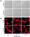
Effect of shear on adherence capacity and on the F-actin of AD-MSCs after 24 h. Representative images of adherent cell cultures (A) and F-actin (red) and nuclei (blue) staining (B) after 5 min or 10 min shearing with different shear rates (1, 2, 2.5 and 3 × 10⁴ s−1). Cell cultures without incipient shearing procedure served as control cultures.

Similar articles

References

    1. Zuk P., Zhu M., Mizuno H., Huang J., Futrell W., Katz A., Benhaim P., Lorenz P., Hedrick M. Multilineage Cells from Human Adipose Tissue: Implications for Cell-Based Therapies. Tissue Eng. 2001;7:211–228. doi: 10.1089/107632701300062859. - DOI - PubMed
    1. Rigotti G., Marchi A., Galiè M., Baroni G., Benati D., Krampera M., Pasini A., Sbarbati A. Clinical treatment of radiotherapy tissue damage by lipoaspirate transplant: A healing process mediated by adipose-derived adult stem cells. Plast. Reconstr. Surg. 2007;119:1409–1422. doi: 10.1097/01.prs.0000256047.47909.71. - DOI - PubMed
    1. Ueberreiter K., Finckenstein J.G.v., Cromme F., Herold C., Tanzella U., Vogt P.M. BEAULI™—eine neue Methode zur einfachen und zuverlässigen Fettzell-Transplantation. Handchir. Mikrochir. Plast. Chir. 2010;42:379–385. doi: 10.1055/s-0030-1267913. - DOI - PubMed
    1. Spiekman M., van Dongen J.A., Willemsen J.C., Hoppe D.L., van der Lei B., Harmsen M.C. The power of fat and its adipose-derived stromal cells: Emerging concepts for fibrotic scar treatment. J. Tissue Eng. Regen. Med. 2017;11:3220–3235. doi: 10.1002/term.2213. - DOI - PMC - PubMed
    1. Stasch T., Hoehne J., Huynh T., Baerdemaeker R.d., Grandel S., Herold C. Débridement and Autologous Lipotransfer for Chronic Ulceration of the Diabetic Foot and Lower Limb Improves Wound Healing. Plast. Reconstr. Surg. 2015;136:1357–1366. doi: 10.1097/PRS.0000000000001819. - DOI - PubMed

LinkOut - more resources