. 2024 Oct 16;14(20):2294.
doi: 10.3390/diagnostics14202294.

Vascular Perspectives of the Midfacial Superficial Musculoaponeurotic System

Affiliations

Vascular Perspectives of the Midfacial Superficial Musculoaponeurotic System

Delia Hînganu et al. Diagnostics (Basel). .

Abstract

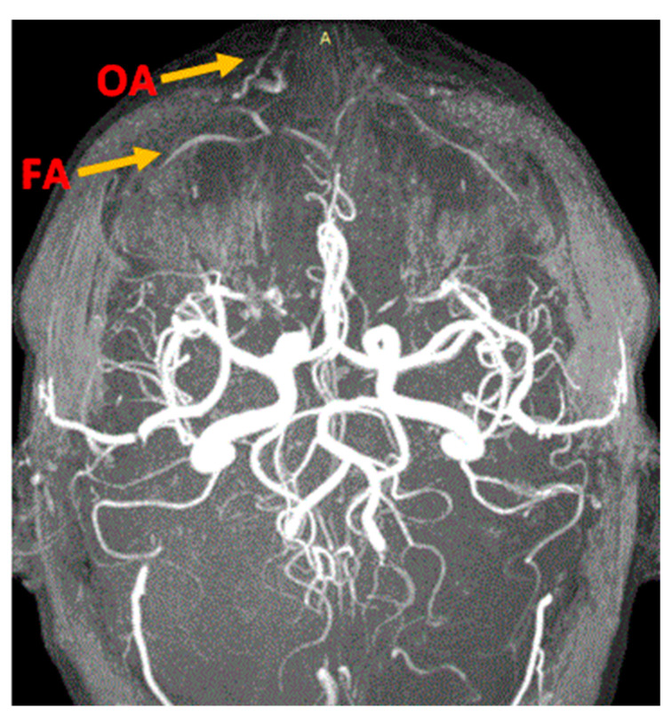
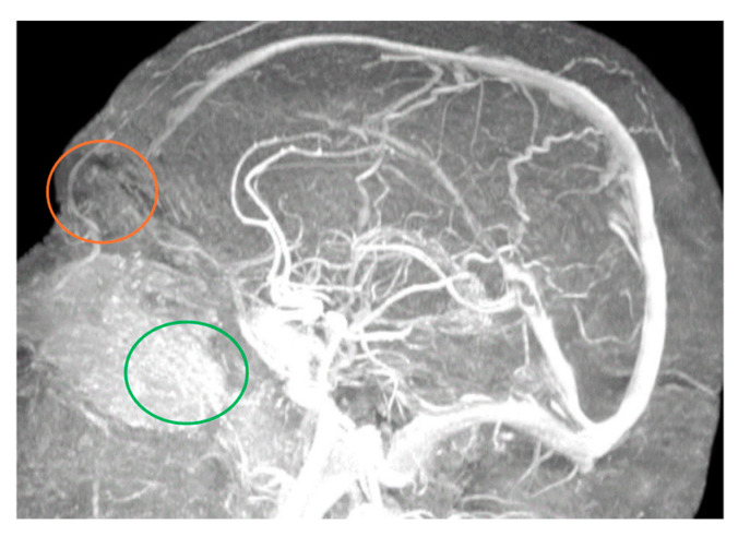
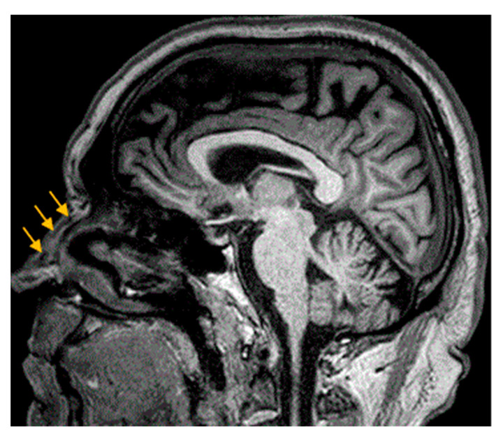
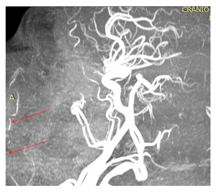
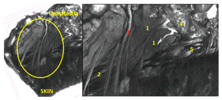
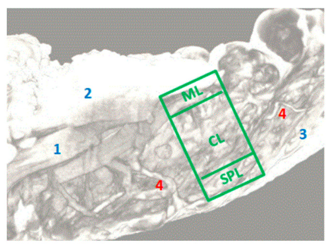
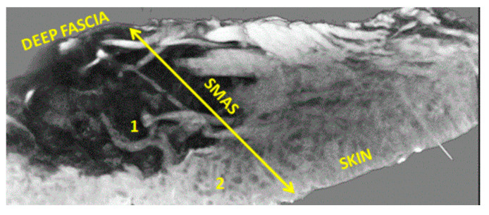
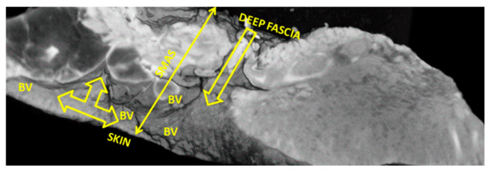
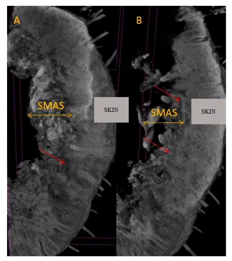
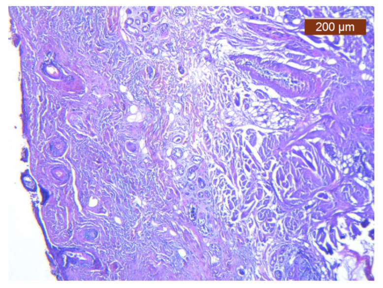
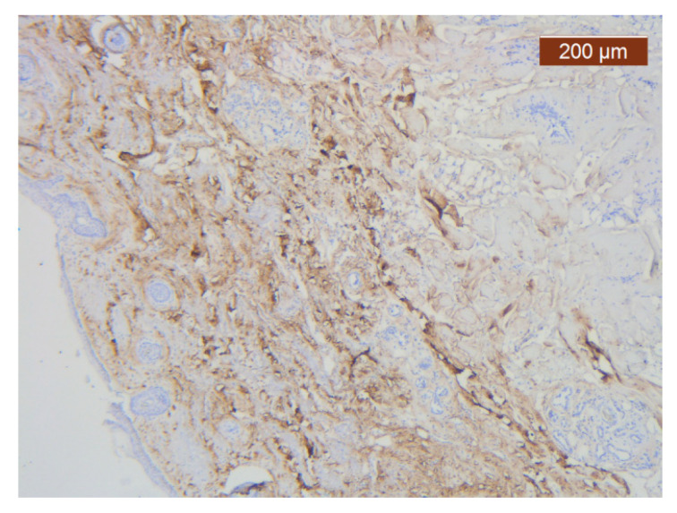
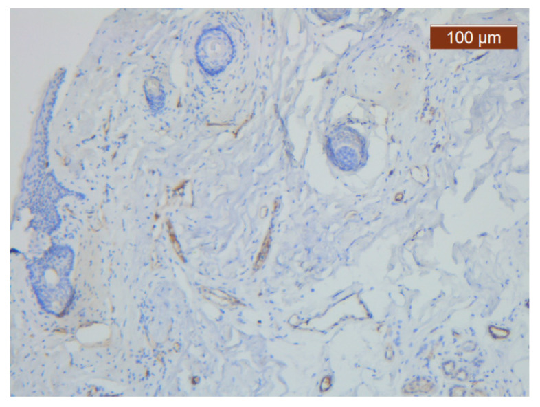
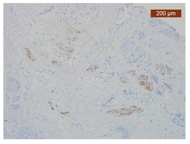
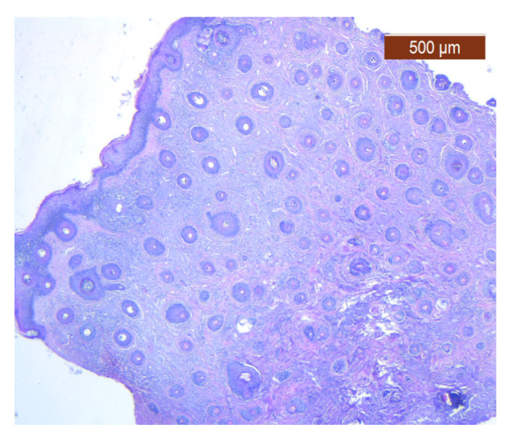
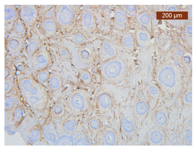
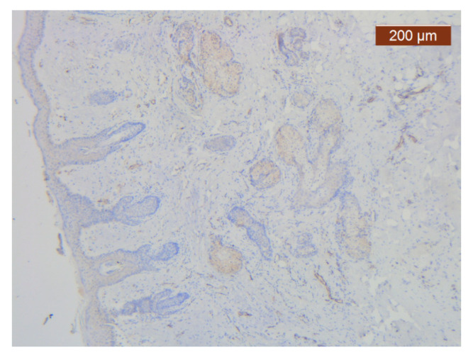
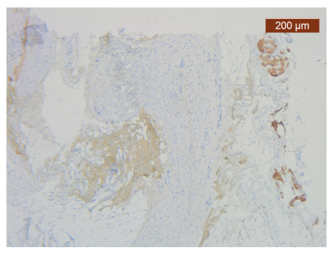
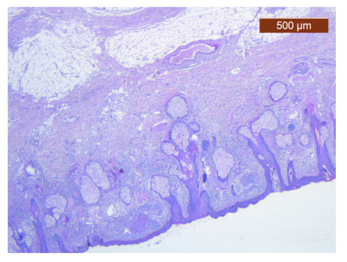
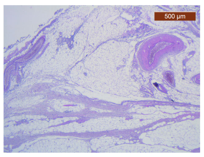
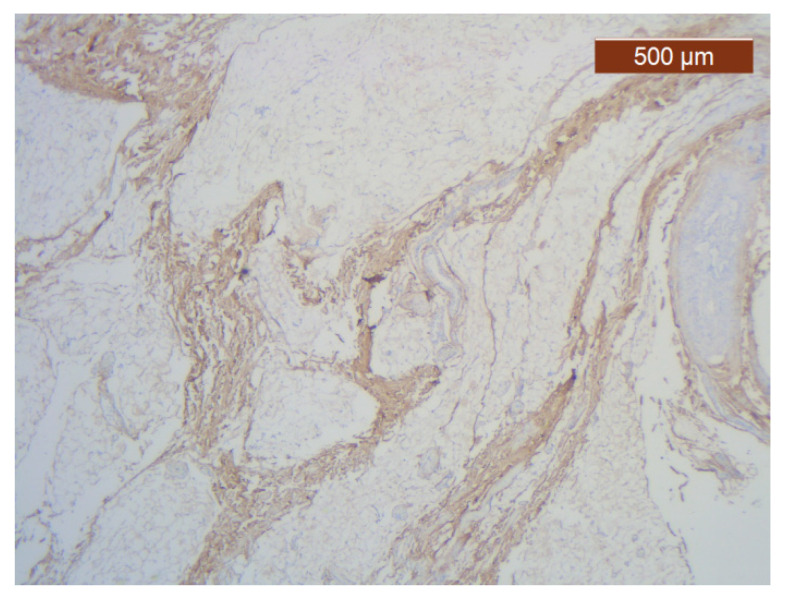
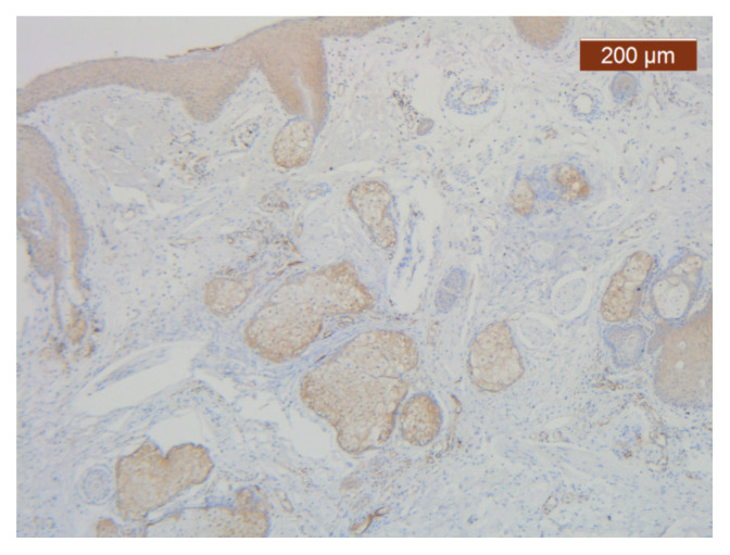
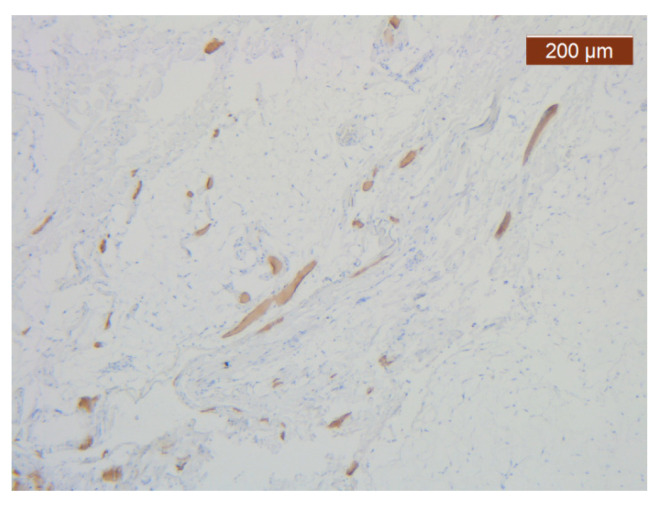
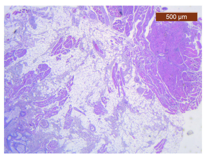
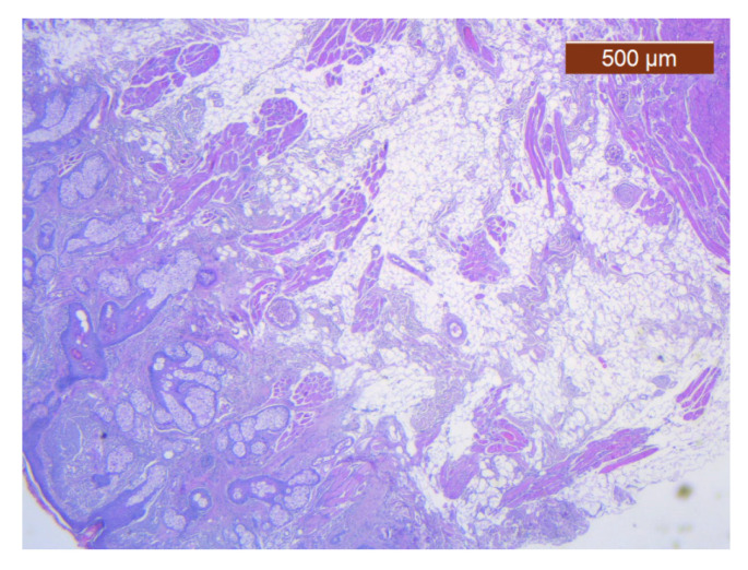
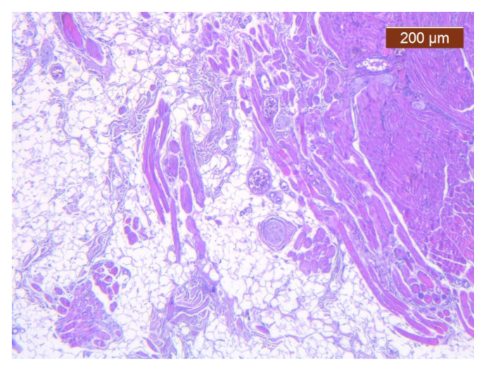
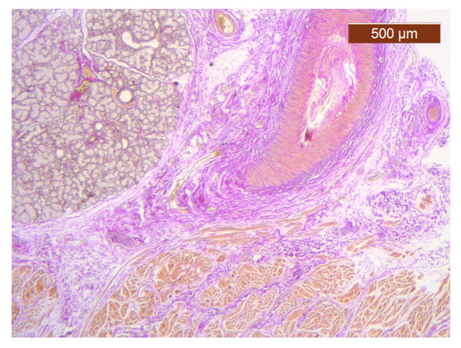
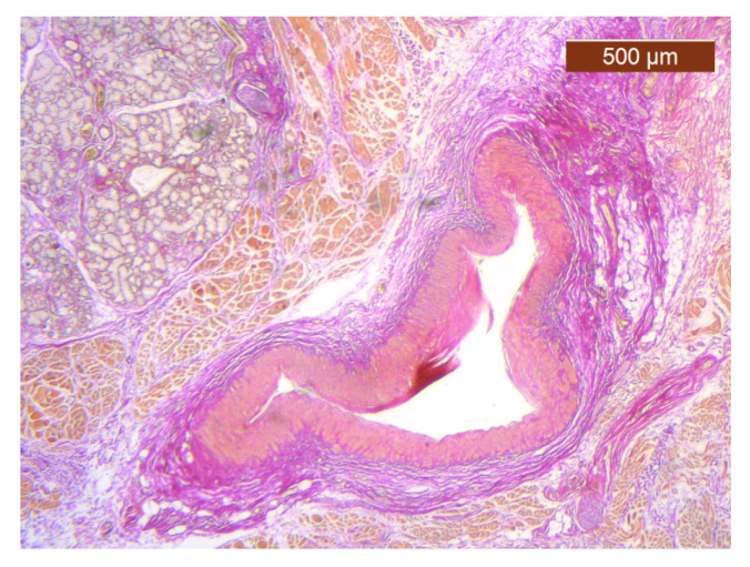
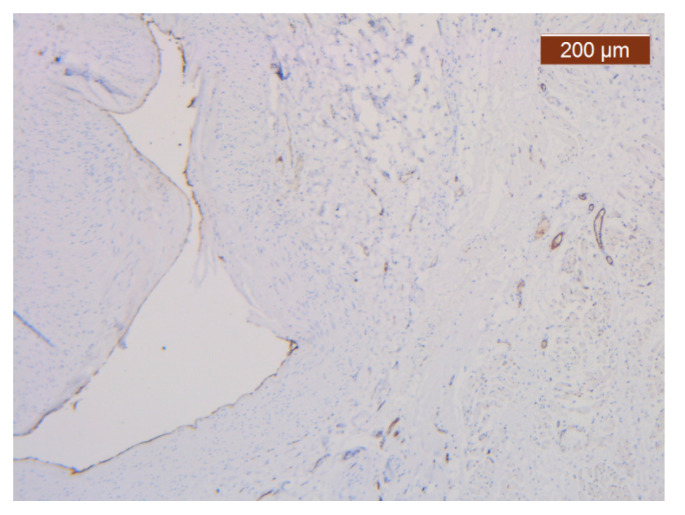
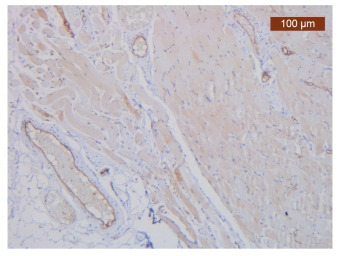
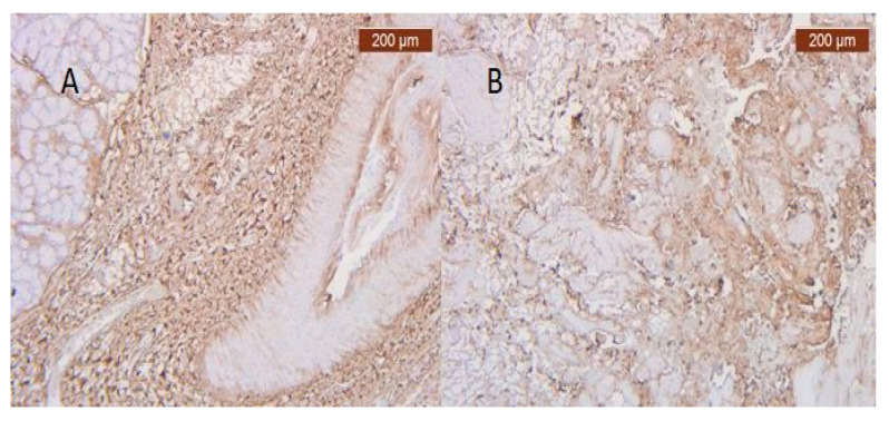
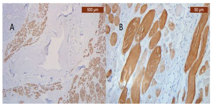
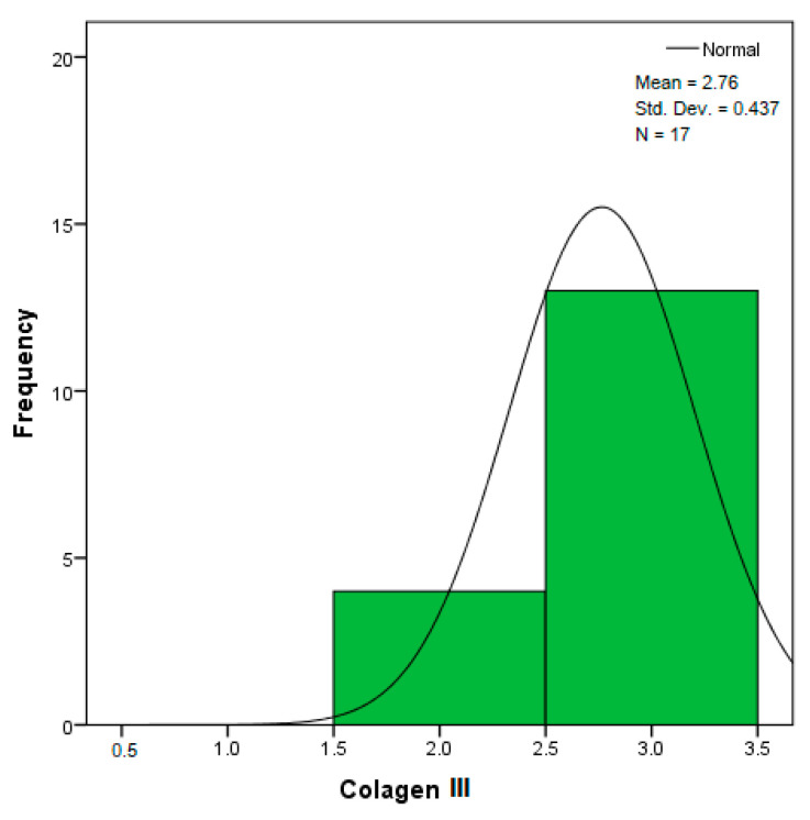
Objectives: Presently, data on the vascularization of the superficial musculoaponeurotic system of the face (SMAS) are lacking. Thus, the present study aimed to provide new conclusive data about the topography, density, and relationship of the SMAS blood vessels with other components, namely, the fibrous connective tissue and muscles. Methods: The study included a control lot of 42 cases from the archive of the radiology department. In this group, nuclear magnetic resonance angiography (MRA) was performed in order to identify the main sources of vascular supply. In the second group, tissue samples were collected from the midfacial region of 45 patients from the Oro-Maxillo-Facial and Plastic and Reconstructive Surgery clinics of 'St. Spiridon' County Clinical Emergency Hospital, Iasi. These patients received surgery for excision of tumoral formations that did not involve SMAS components. These samples underwent micro-CT analysis, hematoxylin and eosin (HE) staining, as well as immunohistochemical (IHC) staining for collagen type III, muscle tissue, and the vascular endothelium. Results: We discovered the particular way in which the SMAS components interrelate with vascularization and the regional differences between them. We have discovered a new vascular network specific to the SMAS, highlighted by both the micro-CT technique and microscopy on slides with special IHC staining. Significant differences were observed in the topographic arrangement, density, and relationships of the microscopic vasculature across midfacial regions. IHC staining provided morphological and functional information about the structure and vascularization of SMAS. Conclusions: The MRA technique could not detect the structural blood vessels of the SMAS and other methods for their in vivo visualization must be sought. The blood vessels of the SMAS mainly follow the topography of the muscle fibers. From the SMAS layer where they are found, the distribution branches reach the stroma of the region and the hypoderm. Our data can contribute to the development of surgical techniques tailored to each individual patient, as well as the enhancement of methods for stimulating cutaneous angiogenesis, improving scarring in this region, and advancing biotissue engineering techniques.

Keywords: MRA; SMAS; collagen; endothelium; vascular microperfusion of face.


Conflict of interest statement

The authors declare no conflicts of interest.

Figures

Figure 1
Figure 1
Branches of facial (FA) and ophthalmic arteries (OA) for the nasal region. MRA VPRV 18 yo healthy patient. 9 min and 13 s time of exposure. A: Artery.
Figure 2
Figure 2
Branches from supraorbital and ophthalmic arteries (orange circle), as well as facial artery terminal nasal branches (green circle). DT 51 yo patient. 12 min and 24 s time of exposure.
Figure 3
Figure 3
Nasal SMAS (orange arrows). TM 68 yo patient. 7 min and 30 s time of exposure.
Figure 4
Figure 4
Identification of the superior (superior red arrow) and inferior (inferior red arrow) labial branches of the facial artery (A).
Figure 5
Figure 5
Specimen immersed in Lugol for 7 days from medial side of nasolabial groove; 7 µm scan; yellow circle = SMAS area; 1 = mimic muscles; 2 = fibrous tissue from superficial fascia; 3 = blood vessel; 4 = fat tissue; 5 = attaching manners.
Figure 6
Figure 6
Specimen immersed in Lugol for 7 days from nasal pyramid area; 1 = mimic muscle (procerus); 2 = sub-SMAS fat tissue; 3 = superficial fascia, 4 = blood vessels; ML = muscle layer; CL = conjunctive layer; SPL = superficial fascia layer; whole green rectangular = SMAS.
Figure 7
Figure 7
Significant proper SMAS blood vessel network (1 and 2) revealed in a specimen immersed in Lugol iodide for 14 days. Micro-CT is still able to distinguish how these vessels protrude into the thick conjunctive tissue under the skin of the tip of the nose.
Figure 8
Figure 8
Single yellow arrow = vasculonervous pathway through SMAS; triple yellow arrow = “T” shape splitting of vasculonervous bundles, with retrograde distribution of SMAS blood vessels; BV = blood vessels.
Figure 9
Figure 9
(A) Micro-CT at upper lip level; (B) micro-CT at the level of the lower lip; SMAS blood vessels are highlighted by red arrows.
Figure 10
Figure 10
Subcutaneous collagen thick septa. Collagen fibers (deep), blood vessels, and fat cells (middle alar area) (HE ×5).
Figure 11
Figure 11
Collagen III is either absent or present in a more irregular distribution (anti-collagen III ×5). Staining intensity: strong.
Figure 12
Figure 12
ICAM-2 staining is confined to internal vascular walls (endothelial cells) (anti-ICAM-2 ×10). Staining intensity: weak to moderate.
Figure 13
Figure 13
Intracytoplasmic MyH2 in muscle cells but not in the surrounding connective tissue (anti-MyH2 ×5). Staining intensity: moderate.
Figure 14
Figure 14
Skin with dense dermis, blood vessels, and some adipose cells (HE ×2.5).
Figure 15
Figure 15
Collagen is plentiful in the stroma (anti-collagen III ×5). Staining intensity: moderate to strong.
Figure 16
Figure 16
ICAM-2 moderate positivity in blood vessels (anti-ICAM-2 ×5). Staining intensity: moderate.
Figure 17
Figure 17
MyH2 showed irregular bundles and inegal staining distribution (MyH2 ×5). Staining intensity: moderate.
Figure 18
Figure 18
Collagen fiber meshwork and a large blood vessel with thick walls and lobules of adipose tissue (HE ×2.5).
Figure 19
Figure 19
Details of collagen fiber meshwork and a large blood vessel with thick walls and lobules of adipose tissue (HE ×2.5).
Figure 20
Figure 20
Abundant perivascuar and interstitial collagen III and around adipose cells in soft part of hypodermis (anti-collagen III ×2.5). Staining intensity: moderate to strong.
Figure 21
Figure 21
ICAM-2 staining was negative except in blood vessel walls (anti-ICAM-2 ×10 (left), 5 (right)). Staining intensity: weak to moderate.
Figure 22
Figure 22
Muscular fibers in deep area of dermis (anti-MyH2 ×5). Staining intensity: moderate.
Figure 23
Figure 23
Fibroadipose tissue over the SMAS, well represented with the average thickness of the soft tissue structure (HE ×2.5). Histological piece from biopsy of the lower lip.
Figure 24
Figure 24
General histological view that showed the depth of the SMAS from the surface of the skin (HE ×2.5). Histological piece from biopsy of the lower lip.
Figure 25
Figure 25
Muscle fibers in the SMAS layer (SMAS fibrous septum). SMAS fibrous septa are arranged perpendicular to the skin and enclose the univacuolar compartments of fat cells (HE ×5). Histological piece from biopsy of the lower lip.
Figure 26
Figure 26
Median layers in the labial region, from top to bottom: salivary glands, skeletal muscle (yellow), and collagen fibers (red) (VG ×2.5). Histological piece from biopsy of the upper lip.
Figure 27
Figure 27
Median layers in the labial region, from top to bottom: salivary glands, skeletal muscle, muscular artery, and perivascular collagen fibers (VG ×2.5). Histological piece from biopsy of the upper lip.
Figure 28
Figure 28
ICAM-2 expression in blood vessel walls and largely absent from the surrounding stroma (anti-ICAM-2 ×5). Color intensity: moderate.
Figure 29
Figure 29
ICAM-2 moderately-intensely expressed in blood vessels of the lower lip (anti-ICAM-2 ×10).
Figure 30
Figure 30
(A) Irregular distributions of type III interstitial collagen around the salivary glands (anti-collagen ×5). Color intensity: strong; (B) Collagen is abundant in the stroma. The distribution appears predominantly pericellular (anti-collagen III ×5).
Figure 31
Figure 31
(A) Reaction for immunohistochemically positive myosin of muscle fibers in the SMAS fibrous zone (anti-MyH2 ×5). Intensity of coloring at the level of the upper lip: moderate; (B) Immunohistochemically positive myosin reaction of muscle fibers in the fibrous area of the SMAS, at the level of the lower lip (anti-MyH2 ×20).
Figure 32
Figure 32
Histogram for the variable collagen III according to the dependent variable nasal anatomical region.
Figure 33
Figure 33
Histogram for the variable collagen III according to the dependent variable oral anatomical region.
Figure 34
Figure 34
Histogram for the variable ICAM-2 according to the dependent variable nasal anatomical region.
Figure 35
Figure 35
Histogram for the variable ICAM-2 according to the dependent variable oral anatomical region.
Figure 36
Figure 36
Histogram for the variable MyoH2 according to the dependent variable nasal anatomical region.
Figure 37
Figure 37
Histogram for the variable MyoH2 according to the dependent variable oral anatomical region.
Figure 38
Figure 38
Histogram for the variable LTD according to the dependent variable nasal anatomical region.
Figure 39
Figure 39
Histogram for the variable LTD according to the dependent variable oral anatomical region.
Figure 40
Figure 40
Histogram for the variable LVD according to the dependent variable nasal anatomical region.
Figure 41
Figure 41
Histogram for the variable LVD according to the dependent variable oral anatomical region.
Figure 42
Figure 42
Histogram for the variable LBV_VHD according to the dependent variable nasal anatomical region.
Figure 43
Figure 43
Histogram for the variable LBV_VHD according to the dependent variable oral anatomical region.

Similar articles

References

    1. Cotofana S., Fratila A.A., Schenck T.L., Redka-Swoboda W., Zilinsky I., Pavicic T. The Anatomy of the Aging Face: A Review. Facial Plast. Surg. 2016;32:253–260. doi: 10.1055/s-0036-1582234. - DOI - PubMed
    1. Hînganu D., Scutariu M.M., Hînganu M.V. The existence of labial SMAS-Anatomical, imaging and histological study. Ann. Anat. 2018;218:271–275. doi: 10.1016/j.aanat.2018.04.009. - DOI - PubMed
    1. Stuzin J.M., Rohrich R.J., Dayan E. The Facial Fat Compartments Revisited: Clinical Relevance to Subcutaneous Dissection and Facial Deflation in Face Lifting. Plast. Reconstr. Surg. 2019;144:1070–1078. doi: 10.1097/PRS.0000000000006181. - DOI - PubMed
    1. Kim C.H. Evaluating the Compartment-Specific Effects in Superficial Facial Fat Compartments After Thread-Lifts by the Tensiometer and FACE-Q. Aesthet. Surg. J. Open Forum. 2022;4:ojac065. doi: 10.1093/asjof/ojac065. - DOI - PMC - PubMed
    1. Letourneau A., Daniel R.K. The Superficial Musculoaponeurotic System of the Nose. Plast. Reconstr. Surg. 1988;82:56–57. doi: 10.1097/00006534-198882010-00011. - DOI - PubMed

LinkOut - more resources