Review
. 2024 Oct 21;14(20):2339.
doi: 10.3390/diagnostics14202339.

Comments and Illustrations of the European Federation of Societies for Ultrasound in Medicine (EFSUMB) Guidelines: Rare Malignant Pulmonal and Pleural Tumors: Primary Pulmonary Sarcoma and Mesothelioma, Imaging Features on Transthoracic Ultrasound

Affiliations
Review

Comments and Illustrations of the European Federation of Societies for Ultrasound in Medicine (EFSUMB) Guidelines: Rare Malignant Pulmonal and Pleural Tumors: Primary Pulmonary Sarcoma and Mesothelioma, Imaging Features on Transthoracic Ultrasound

Kathleen Möller et al. Diagnostics (Basel). .

Abstract

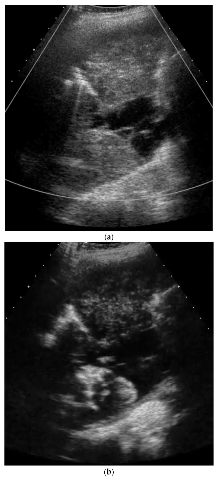
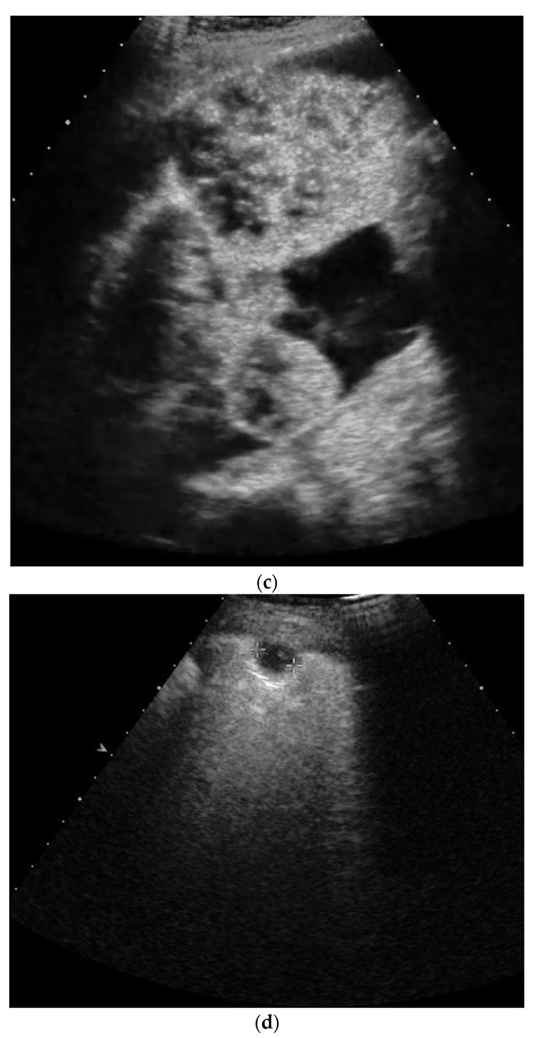
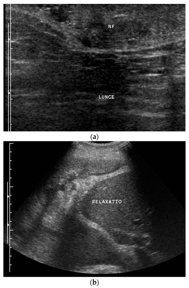
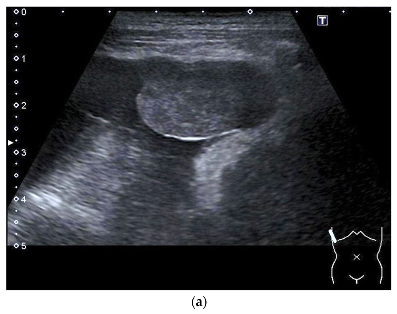
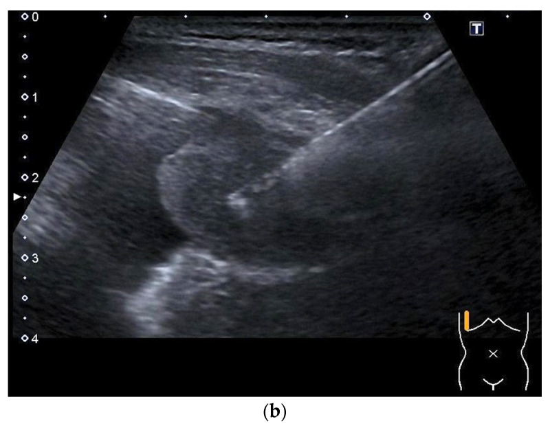
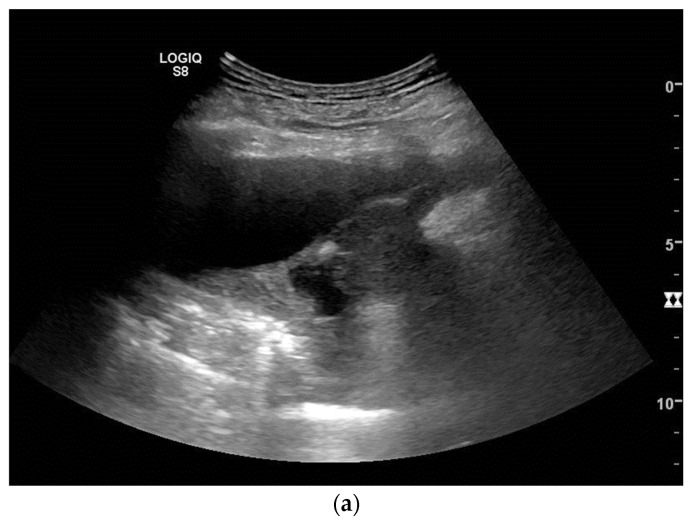
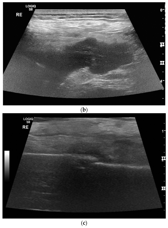
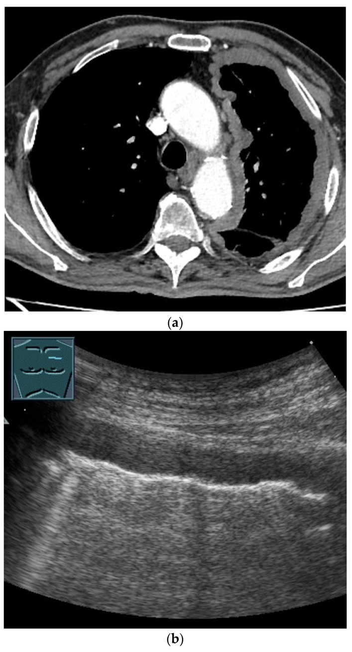
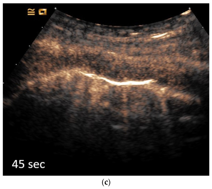
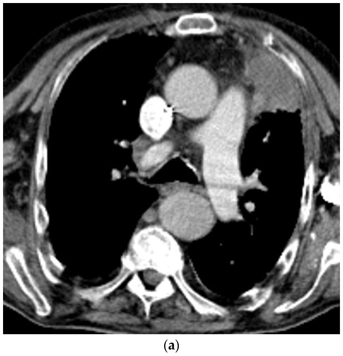
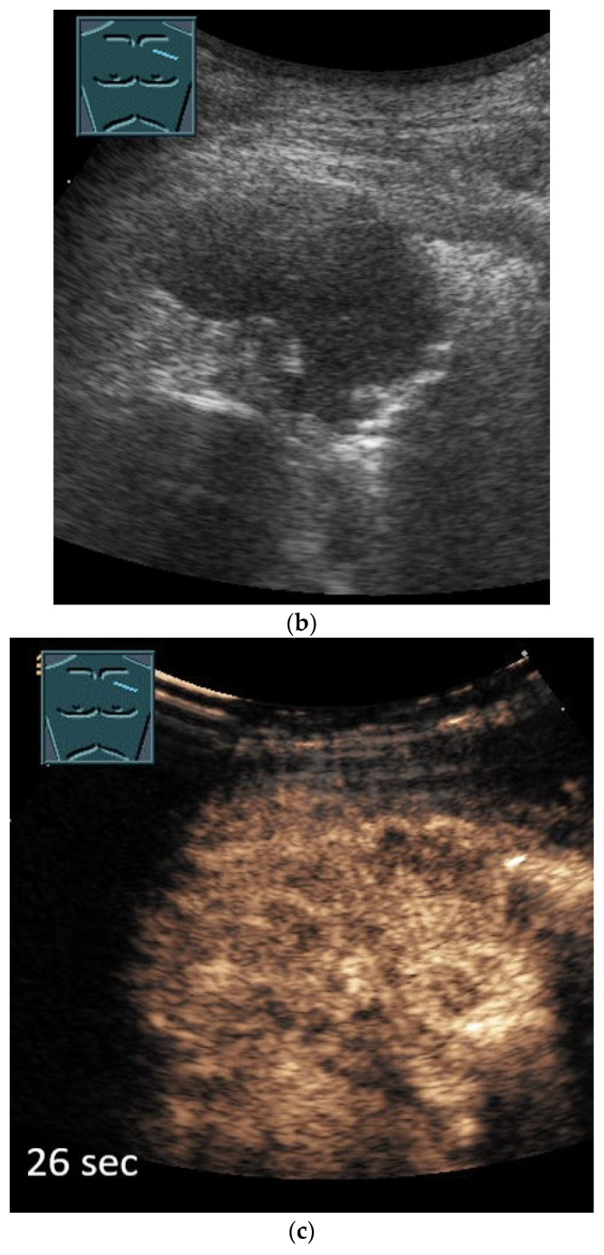
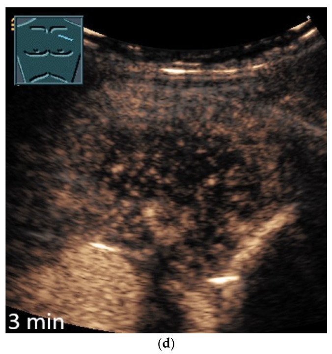
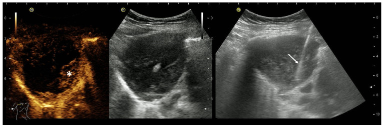
Primary pulmonary sarcoma and mesothelioma are rare malignancies. The review article discusses the appearance of these tumors in B-mode ultrasound (US), color Doppler ultrasound and contrast-enhanced ultrasound (CEUS). In particular, the article is intended to inspire the examination of thoracic wall tumors and pleural masses with the possibilities of ultrasonography and to obtain histologically evaluable material using US or CEUS-guided sampling.

Keywords: advances; contrast-enhanced ultrasound; diagnosis; imaging; lung ultrasound; respiratory medicine.


Conflict of interest statement

The authors declare that they have no financial conflict of interest with regard to the content of this report. Some authors have received financial support and/or honoraria from Bracco for the organization of ultrasound courses. In addition, some authors have been supported with equipment from various ultrasound equipment companies for the organization of ultrasound courses and/or have received honoraria for lectures.

Figures

Figure 1
Figure 1
Primary sarcoma of the lung infiltrating almost the entire hemithorax proven by ultrasound-guided biopsy and histological evaluation with pleural metastasis. B mode ultrasound does not allow a clear differentiation between the lung parenchyma and the tumor (a). Contrast-enhanced ultrasound facilitates potential differentiation, because the atelectatic lung parenchyma is enhancing early via the pulmonary artery circulation (b), whereas the sarcoma shows later centripetal enhancement and non-enhancing (necrotic) areas (c). Multiple pleural metastases as well as a small pleural metastasis were found (d).
Figure 1
Figure 1
Primary sarcoma of the lung infiltrating almost the entire hemithorax proven by ultrasound-guided biopsy and histological evaluation with pleural metastasis. B mode ultrasound does not allow a clear differentiation between the lung parenchyma and the tumor (a). Contrast-enhanced ultrasound facilitates potential differentiation, because the atelectatic lung parenchyma is enhancing early via the pulmonary artery circulation (b), whereas the sarcoma shows later centripetal enhancement and non-enhancing (necrotic) areas (c). Multiple pleural metastases as well as a small pleural metastasis were found (d).
Figure 2
Figure 2
Potential neurofibroma of the thoracic wall (a) and biopsy-proven neurofibrosarcoma of the lung infiltrating almost the entire hemithorax (left and therefore upper part of the image) with paralysis of the diaphragm and bulging of the liver into the thorax (“Relaxatio”) (b) in a patient with neurofibromatosis type 1 disease. Lunge: Lung. The origin of the sarcoma remained unresolved. Neurofibromas were also evident in the skin and abdomen.
Figure 3
Figure 3
Mesothelioma (epithelioid cell type) in an 81-year-old female patient hospitalized for shortness of breath, vertigo and a weight loss of 10 kg in 6 months. There was a history of breast cancer 15 years ago with complete remission after surgery and adjuvant radiation, but no history of known exposure to asbestos. Ultrasound revealed a large unilateral pleural effusion with a solid tumor at the parietal pleura in the costodiaphragmal recess (a). Histology from biopsies confirmed mesothelioma (b).
Figure 3
Figure 3
Mesothelioma (epithelioid cell type) in an 81-year-old female patient hospitalized for shortness of breath, vertigo and a weight loss of 10 kg in 6 months. There was a history of breast cancer 15 years ago with complete remission after surgery and adjuvant radiation, but no history of known exposure to asbestos. Ultrasound revealed a large unilateral pleural effusion with a solid tumor at the parietal pleura in the costodiaphragmal recess (a). Histology from biopsies confirmed mesothelioma (b).
Figure 4
Figure 4
Year-old male patient hospitalized for shortness of breath and located right-sided chest pain. There was no history of asbestos exposure. Ultrasound revealed pleural effusion and tumor masses at the parietal pleura (a) with infiltration of the diaphragm and chest wall (b) thereby causing a rib fracture (c). Histology from biopsies confirmed mesothelioma.
Figure 4
Figure 4
Year-old male patient hospitalized for shortness of breath and located right-sided chest pain. There was no history of asbestos exposure. Ultrasound revealed pleural effusion and tumor masses at the parietal pleura (a) with infiltration of the diaphragm and chest wall (b) thereby causing a rib fracture (c). Histology from biopsies confirmed mesothelioma.
Figure 5
Figure 5
Eighty-year-old patient with a history of asbestos exposure and histologically confirmed epithelioid mesothelioma illustrated on CT (a) and B-mode US (b). The extensive pleural thickening forming a circular wall around the left lung can be seen on CT (a). B-mode US shows a clear hypoechoic thickening of the pleura, which was measured at more than 10 mm (b). The lesion is characterized by a moderate systemic arterial enhancement on CEUS (c).
Figure 5
Figure 5
Eighty-year-old patient with a history of asbestos exposure and histologically confirmed epithelioid mesothelioma illustrated on CT (a) and B-mode US (b). The extensive pleural thickening forming a circular wall around the left lung can be seen on CT (a). B-mode US shows a clear hypoechoic thickening of the pleura, which was measured at more than 10 mm (b). The lesion is characterized by a moderate systemic arterial enhancement on CEUS (c).
Figure 6
Figure 6
Eighty-year-old patient with histologically confirmed epithelioid mesothelioma shown on CT (a) and B-Mode ultrasound (b). On CEUS, the lesion shows a marked bronchial arterial enhancement (26 s) (c) with parenchymal washout (d).
Figure 6
Figure 6
Eighty-year-old patient with histologically confirmed epithelioid mesothelioma shown on CT (a) and B-Mode ultrasound (b). On CEUS, the lesion shows a marked bronchial arterial enhancement (26 s) (c) with parenchymal washout (d).
Figure 6
Figure 6
Eighty-year-old patient with histologically confirmed epithelioid mesothelioma shown on CT (a) and B-Mode ultrasound (b). On CEUS, the lesion shows a marked bronchial arterial enhancement (26 s) (c) with parenchymal washout (d).
Figure 7
Figure 7
CEUS-guided biopsy of a subpleural lung tumor of a 66-year-old female patient. CEUS revealed large non-enhancing areas; only a small peripheral area was enhancing in the arterial phase (*). The tip of the biopsy needle (arrow) was positioned in this vital part of the tumor to facilitate the procurement of an adequate sample.
Figure 8
Figure 8
A male patient reported asbestos exposure about 45 years ago. CT imaging revealed a tumor in the mediastinal pleura of the right upper thorax (a,b). EUS performed transesophageally with an EBUS bronchoscope (Olympus) (EUS-B) showed a solid inhomogeneous tumor and adjacent compressed lung tissue (c). The performance of an EUS-B-FNB (Mediglobe Top-Gain 22 G) confirmed an epithelioid mesothelioma (d).
Figure 8
Figure 8
A male patient reported asbestos exposure about 45 years ago. CT imaging revealed a tumor in the mediastinal pleura of the right upper thorax (a,b). EUS performed transesophageally with an EBUS bronchoscope (Olympus) (EUS-B) showed a solid inhomogeneous tumor and adjacent compressed lung tissue (c). The performance of an EUS-B-FNB (Mediglobe Top-Gain 22 G) confirmed an epithelioid mesothelioma (d).

Similar articles

Cited by

References

    1. Dietrich C.F., Gorg C., Horn R., Prosch H., Safai Zadeh E., Jenssen C. Ultrasound of the lung. Ultraschall Med. 2023;44:582–599. doi: 10.1055/a-2010-7282. - DOI - PubMed
    1. Safai Zadeh E., Gorg C., Prosch H., Horn R., Jenssen C., Dietrich C.F. The Role of Thoracic Ultrasound for Diagnosis of Diseases of the Chest Wall, the Mediastinum, and the Diaphragm-Narrative Review and Pictorial Essay. Diagnostics. 2023;13:767. doi: 10.3390/diagnostics13040767. - DOI - PMC - PubMed
    1. Horn R., Gorg C., Prosch H., Safai Zadeh E., Jenssen C., Dietrich C.F. Sonography of the pleura. Ultraschall Med. 2024;45:118–146. doi: 10.1055/a-2189-5050. - DOI - PubMed
    1. Beshara M., Bittner E.A., Goffi A., Berra L., Chang M.G. Nuts and bolts of lung ultrasound: Utility, scanning techniques, protocols, and findings in common pathologies. Crit. Care. 2024;28:328. doi: 10.1186/s13054-024-05102-y. - DOI - PMC - PubMed
    1. Fang C., Jaworska J., Buda N., Ciuca I.M., Dong Y., Feldkamp A., Jungert J., Kosiak W., Mentzel H.J., Pienar C., et al. Ultrasound of the chest and mediastinum in children, interventions and artefacts. WFUMB review paper (part 3) Med. Ultrason. 2022;24:65–67. doi: 10.11152/mu-3323. - DOI - PubMed