. 2024 Oct 6;14(10):479.
doi: 10.3390/bios14100479.

Comparison of Survivin Determination by Surface-Enhanced Fluorescence and Raman Spectroscopy on Nanostructured Silver Substrates

Affiliations

Comparison of Survivin Determination by Surface-Enhanced Fluorescence and Raman Spectroscopy on Nanostructured Silver Substrates

Georgia Geka et al. Biosensors (Basel). .

Abstract

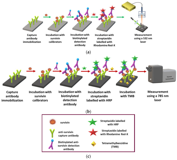
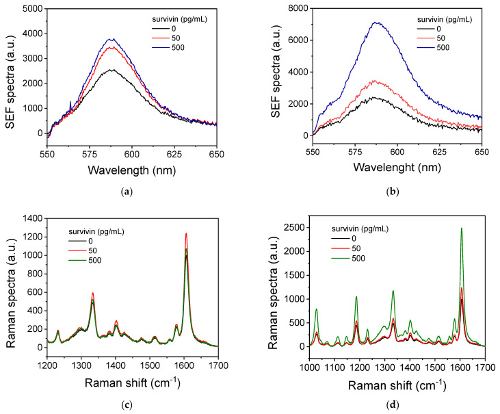
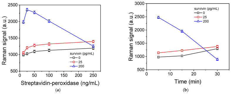
Survivin belongs to a family of proteins that promote cellular proliferation and inhibit cellular apoptosis. Its overexpression in various cancer types has led to its recognition as an important marker for cancer diagnosis and treatment. In this work, we compare two approaches for the immunochemical detection of survivin through surface-enhanced fluorescence or Raman spectroscopy using surfaces with nanowires decorated with silver nanoparticles in the form of dendrites or aggregates as immunoassays substrates. In both substrates, a two-step non-competitive immunoassay was developed using a pair of specific monoclonal antibodies, one for detection and the other for capture. The detection antibody was biotinylated and combined with streptavidin labeled with rhodamine for the detection of surface-enhanced fluorescence, while, for the detection via Raman spectroscopy, streptavidin labeled with peroxidase was used and the signal was obtained after the application of 3,3',5,5'-tetramethylbenzidine (TMB) precipitating substrate. It was found that the substrate with the silver dendrites provided higher fluorescence signal intensity compared to the substrate with the silver aggregates, while the opposite was observed for the Raman signal. Thus, the best substrate was used for each detection method. A detection limit of 12.5 pg/mL was achieved with both detection approaches along with a linear dynamic range up to 500 pg/mL, enabling survivin determination in human serum samples from both healthy and ovarian cancer patients for cancer diagnosis and monitoring purposes.

Keywords: cancer marker; immunochemical detection; optical biosensor; surface-enhanced Raman spectroscopy; surface-enhanced photoluminescence; survivin.


Conflict of interest statement

The authors declare no conflict of interest.

Figures

Figure 1
Figure 1
Schematic illustration of the immunoassay for survivin determination with SEF (a) or SERS (b) and of the reagents used in both assays (c).
Figure 2
Figure 2
Characteristic cross and top view SEM images of Si nanowires with height of approximately 250 nm decorated with 800 nm-long Ag dendrites (a,b) or Si nanowires with height of approximately 500 nm decorated with approximately 150 nm-long Ag aggregates (c,d). The cross-view images (a,c) have been received with a higher magnification to show in detail the Ag nanoparticles’ structure whereas the top view images (b,d) with a lower magnification to demonstrate the distribution of these structures across the substrate surface.
Figure 3
Figure 3
(a,b) SEF spectra obtained from nanostructured Ag/Si substrates with Ag dendrites following a one-step (a) or a two-step survivin immunoassay (b) for the zero calibrator (black line) and calibrators containing 50 (red line) and 500 pg/mL (blue line). (c,d) SERS spectra obtained from nanostructured Ag/Si substrates with Ag aggregates following a one-step (c) or a two-step survivin immunoassay (d) for the zero calibrator (black line) and calibrators containing 50 (red line) and 500 pg/mL (green line).
Figure 4
Figure 4
(a) SEF signal values corresponding to zero calibrator (plain orange and green columns) and a calibrator containing 50 pg/mL of survivin (striped orange and green columns) obtained using the detection antibody at concentrations of 1.25 (orange columns) and 2.5 μg/mL (green columns). Each point is the mean of three samples ± SD. (b) Survivin calibration curves obtained from nanostructured Ag/Si substrates with Ag dendrites for duration of each immunoassay step equal to 0.5 (squares), 1 (circles), or 2 h (triangles). Each point is the mean of three samples ± SD.
Figure 5
Figure 5
(a) Raman signals corresponding to peak at 1607 cm−1 obtained for the zero calibrator (squares) or calibrators containing 25 (circles) or 200 pg/mL (triangles) of survivin with respect to the streptavidin-peroxidase concentration. The incubation with the precipitating TMB substrate was 15 min. Each point is the mean of three samples ± SD. (b) Raman signals corresponding to peak at 1607 cm−1 obtained for the zero calibrator (squares) or calibrators containing 25 (circles) or 200 pg/mL (triangles) of survivin with respect to the duration of incubation with the precipitating TMB substrate. The streptavidin-peroxidase concentration was 25 ng/mL. Each point is the mean of three samples ± SD.
Figure 6
Figure 6
(a) SEF spectra received from Ag/Si substrates with dendrites for survivin calibrators from 0 to 500 pg/mL in assay buffer. (b) Survivin SEF calibration curve. (c) SERS spectra received from Ag/Si substrates with aggregates for survivin calibrators from 0 to 500 pg/mL in assay buffer. (d) Survivin SERS calibration curve. Each point is the mean value of 10 measurements from 3 replicate samples ± 3SD.

Similar articles

References

    1. Li Y., Lu W., Yang J., Edwards M., Jiang S. Survivin as a biological biomarker for diagnosis and therapy. Expert Opin. Biol. Ther. 2021;21:1429–1441. doi: 10.1080/14712598.2021.1918672. - DOI - PubMed
    1. Stobiecka M., Ratajczak K., Jakiela S. Toward early cancer detection: Focus on biosensing systems and biosensors for an anti-apoptotic protein survivin and survivin mRNA. Biosens. Bioelectron. 2019;137:58–71. doi: 10.1016/j.bios.2019.04.060. - DOI - PubMed
    1. Mahmoudian-Sani M.R., Alghasi A., Saeedi-Boroujeni A., Jalali A., Jamshidi M., Khodadadi A. Survivin as a diagnostic and therapeutic marker for thyroid cancer. Pathol. Res. Pract. 2019;215:619–625. doi: 10.1016/j.prp.2019.01.025. - DOI - PubMed
    1. Santarelli A., Mascitti M., Lo Russo L., Colella G., Giannatempo G., Bambini F., Emanuelli M., Procaccini M., Lo Muzio L. Detection level of salivary survivin in patients with OSCC. J. Carcinog. Mutagen. 2013;S5:4. doi: 10.4172/2157-2518.S5-004. - DOI
    1. Meenakshi Warrier N., Agarwal P., Kumar P. Emerging importance of survivin in stem cells and cancer: The development of new cancer therapeutics. Stem Cell Rev. Rep. 2020;16:828–852. doi: 10.1007/s12015-020-09995-4. - DOI - PMC - PubMed

LinkOut - more resources