Case Reports
. 2024 Sep 27;14(5):2013-2026.
doi: 10.3390/clinpract14050159.

Aesthetical and Functional Rehabilitation for an Ankylosed Maxillary Canine-A Case Report

Affiliations
Case Reports

Aesthetical and Functional Rehabilitation for an Ankylosed Maxillary Canine-A Case Report

Tatiana Roman et al. Clin Pract. .

Abstract

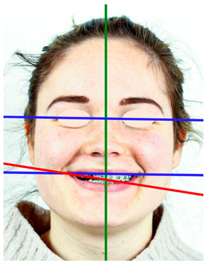
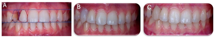
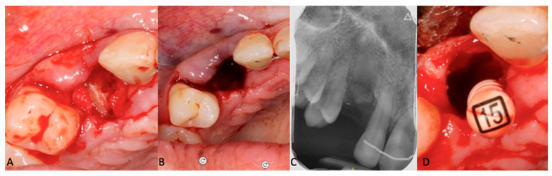
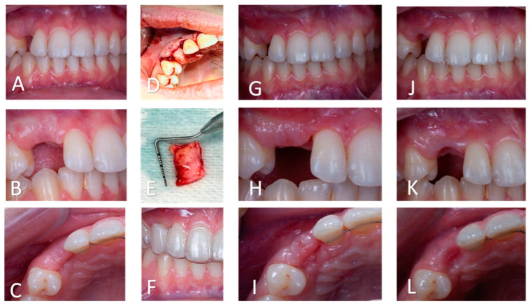
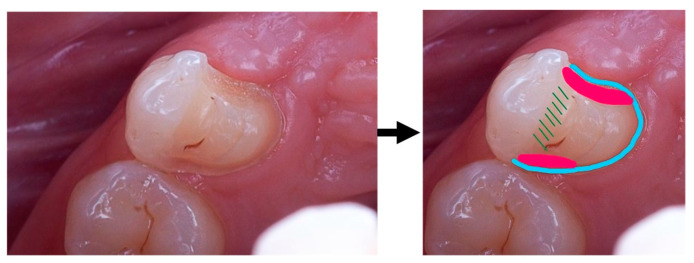
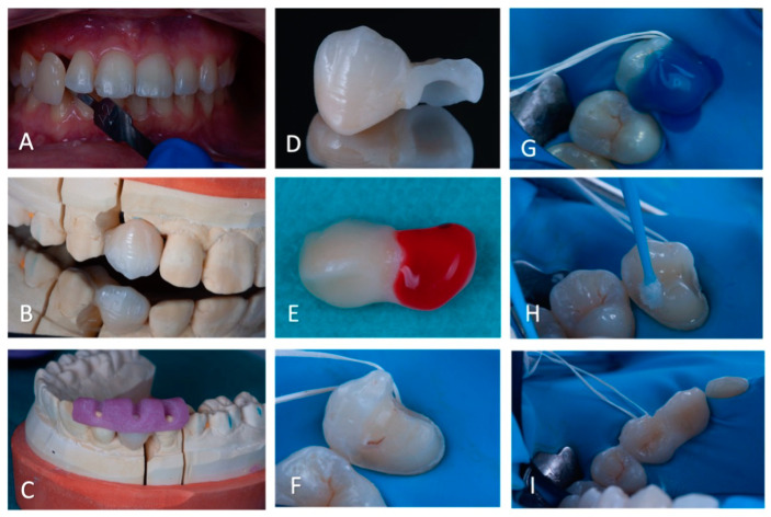
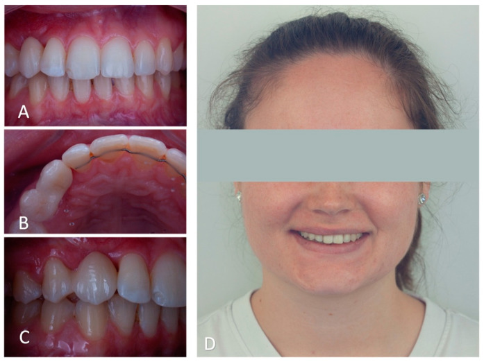
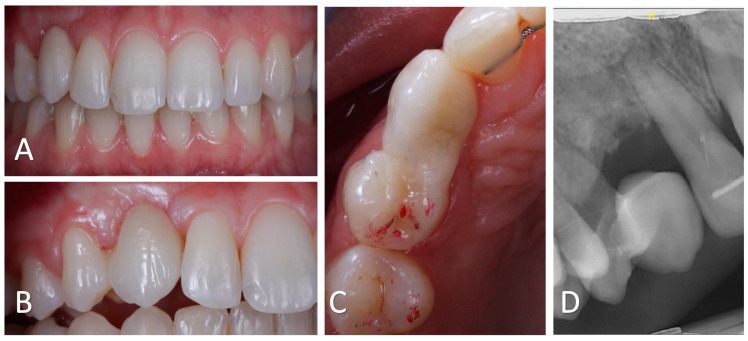
Background: As the functional and aesthetical importance of the canine cannot be overstated, the management of a missing canine is challenging. This case report describes the treatment of an infra-occluded ankylosed maxillary canine in a patient with previously failed orthodontic treatment. Case description: A 20-year-old patient sought a second opinion for orthodontic treatment failure. The patient presented with an impacted, ankylosed, and severely infra-occluded right maxillary canine, as well as an iatrogenic clockwise cant of the maxillary occlusal plane and several root resorptions. The treatment corrected the cant of the occlusal plane while avoiding further root resorption, partially extracted the upper right canine, improved the quality and quantity of the soft tissue in the newly edentulous area, and provided a prosthetic rehabilitation using a lithium disilicate ceramic resin-bonded cantilever bridge. Conclusions: The use of a cantilevered bridge resulted in an aesthetically pleasing and minimally invasive rehabilitation. This technique is reversible, does not affect pulp vitality, and is a viable solution for rehabilitating the smiles of young patients. Clinical significance: The smile rehabilitation for an ankylosed maxillary canine, especially in the case of a previously failed orthodontic treatment, is an important clinical challenge. A minimally invasive long-term restoration with a cantilever bridge is a viable solution. Functional and aesthetically pleasant results can be achieved with a multidisciplinary approach.

Keywords: ankylosed tooth; canine ankylosis; cantilever bridge; case report; decoronation; occlusal plane cant; root resorption.


Conflict of interest statement

The authors declare no conflicts of interest.

Figures

Figure 1
Figure 1
Extra-oral photography of the patient, at the initial appointment with aesthetic analysis. The interpupillary and inter-commissure lines are parallel but not perpendicular to the midsagittal plane; the frontal aesthetic plane is unsightly and presents a clockwise cant. The smile line is low.
Figure 2
Figure 2
Three-dimensional imaging, CBCT (cone beam computed tomography): (A): sagittal section of tooth 13. Thin buccal and palatal cortical bone plates and proximity of the apex of 13 with the right nasal cavity can be noted. (B) Two-dimensional reconstruction of the upper jaw. Important root resorptions of the central maxillary incisors can be noted. A root resorption of the lateral incisor is also present.
Figure 3
Figure 3
Frontal intraoral views in occlusion, right lateral views, occlusal views of the maxillary arch: (AC)—initial situation, at the first consultation; (DF)—intermediate situation, during the second orthodontic treatment; (GI)—clinical situation upon debonding, (J)—post-treatment orthopantomogram.
Figure 4
Figure 4
Smile Design. In white, shape of # 23 in position of # 13: the mesio-distal space available for the # 13 tooth is considerably larger than the mesiodistal size of # 23. In lilac, gingival and cervical embrasure spaces: there are considerable discrepancies in the embrasures’ form in the vicinity of # 13. In green, gingival zenith of the anterior teeth showing considerable alignment discrepancies. In red, aesthetic plane, clockwise canted.
Figure 5
Figure 5
(A): Virtual aesthetic project (Smile Design). In black: the current size of tooth # 12 and projection of tooth # 13 with the same dimensions as tooth # 23. In red: Modification of the shape of tooth # 12, optimising the shape of the occlusal and cervical embrasure between tooth # 12 and tooth # 13 and bringing the incisal edge of tooth # 12 back into a harmonious aesthetic plane. In blue: Proposed enlarged shape for tooth # 13 to optimise the shape of the cervical embrasure. (B): Mock-up with replacement of tooth # 13 (enlarged) and modification of the shape of tooth # 12. (C): Mock-up without modification of the shape of tooth # 12, but with enlarged shape of # 13.
Figure 6
Figure 6
(AC): Pre-operative view of the decoronation. (C): Post-operative intraoral teleradiograph. (D): Pre-operative view of the supra-radicular blood clot formation induced by the remaining pulp parenchyma from the apical fragment.
Figure 7
Figure 7
Soft tissue management. (AC): Intra-oral views (frontal, lateral, and occlusal, respectively) after decoronation and before periodontal surgery. The soft tissues are thin and the gingival lines of # 13 and # 23 are asymmetric. (D): Intra-oral pre-operative view of the mucogingival surgery. Buccal and supra-crestal placement of the graft. (E): Connective tissue graft. (F): Intra-oral frontal view with the vacuum-formed retainer, worn during healing. (GI): Intra-oral views (frontal, lateral, and occlusal, respectively) after periodontal surgery. An increase in the soft tissue thickness in the frontal and transversal planes is observed. (JL): Intra-oral views (frontal, lateral, and occlusal, respectively) after gingival modelling with the retainer and the veneer tooth. The gingival line is aesthetically pleasing.
Figure 8
Figure 8
Cantilever bridge retainer design: blue—proximal and palatal chamfer; red—proximal boxes, with a larger box facing the edentulous region; green—homothetic reduction in the palatal cusp.
Figure 9
Figure 9
(A): Intra-oral photography displaying the visual colour assessment, using a shade-guide Table (B): Buccal view of the bridge positioned on the plaster model. (C): Physical plaster model with the resin repositioning key. (D): Buccal view of the cantilever bridge displaying the thickness of the retainer wing. (E): Etching of the intrados of the bridge. (F): Placement of the surgical field. (G): Etching of the supporting tooth. (H): Application of adhesive on the supporting tooth # 14. (I): Cantilever bridge immediately after bonding displaying the fit on the supporting tooth.
Figure 10
Figure 10
Immediate post-operatory images. (A): Intra-oral frontal view shows the black triangle between # 12 and # 13. (B): Occlusal view shows palatal cusp coverage of # 14 and the size of the connector. (C): Lateral intra-oral view of the bonded tooth # 13 displaying the gingival integration of the freshly bonded bridge. (D): Extra-oral frontal view of the patient’s smile, displaying the aesthetic integration of the cantilever bridge.
Figure 11
Figure 11
Follow-up at 19 months after surgery. (A): Intra-oral frontal view showing satisfactory aesthetics of the bonded bridge. The black triangle between # 13 and # 12 appears to be smaller. (B). Intra-oral lateral view displaying the healed gingiva in the cervical embrasure on both to the cantilever bridge. (C): Occlusal view of # 13 showing the absence of static or dynamic occlusal charge. (D): Sagittal CBCT radiograph showing bone preservation and partial replacement of the residual root by bone.

Similar articles

Cited by

References

    1. Becker A., Chaushu S. Etiology of maxillary canine impaction: A review. Am. J. Orthod. Dentofacial Orthop. 2015;148:557–567. doi: 10.1016/j.ajodo.2015.06.013. - DOI - PubMed
    1. Garcia A. Ankylosis of impacted canines: A retrospective post-surgical study. Int. Orthod. 2023;11:422–431. doi: 10.1016/j.ortho.2013.09.009. - DOI - PubMed
    1. Parrish J., Hills L., Ganesan K. Management of palatally ectopic canines part 2: Ankylosis hypotheses and southend experience. Adv. Mech. Mach. Sci. 2022;8:100349. doi: 10.1016/j.adoms.2022.100349. - DOI
    1. Rodrigues D.B., Wolford L.M., Figueiredo L.M.G., Adams G.Q. Management of Ankylosed Maxillary Canine with Single-Tooth Osteotomy in Conjunction with Orthognathic Surgery. J. Oral Maxillofac. Surg. 2014;72:2419.e1-6. doi: 10.1016/j.joms.2014.07.016. - DOI - PubMed
    1. Oliveira M.V., Pithon M.M. Attempted traction of impacted and ankylosed maxillary canines. Am. J. Orthod. Dentofac. Orthop. 2012;142:106–114. doi: 10.1016/j.ajodo.2010.09.037. - DOI - PubMed

Publication types

LinkOut - more resources