

A Single-Centre Analysis of Surgical Techniques for Myelomeningocele Closure: Methods, Outcomes, and Complications
- PMID: 39451877
- PMCID: PMC11506740
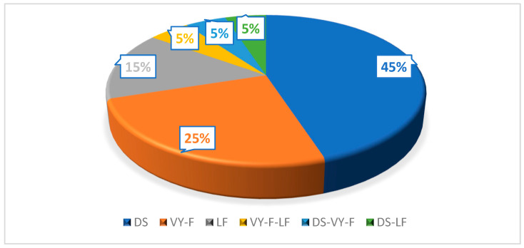
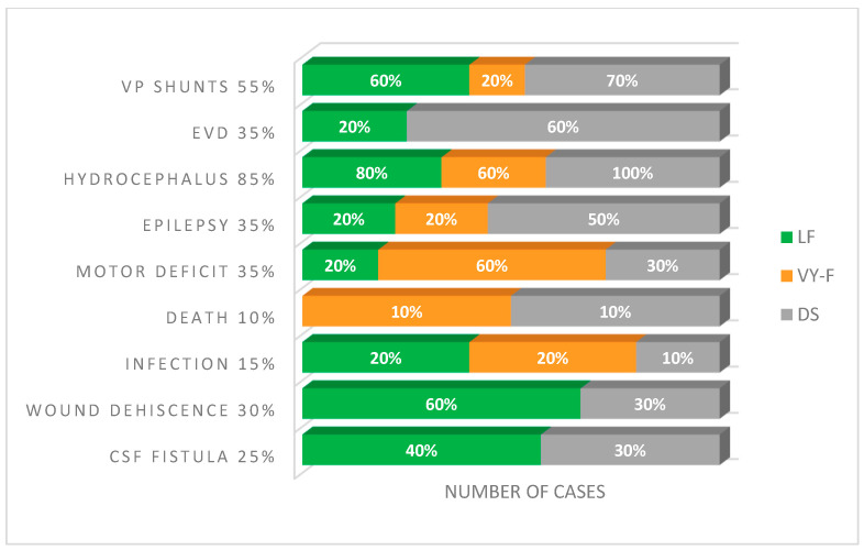
- DOI: 10.3390/clinpract14050162
A Single-Centre Analysis of Surgical Techniques for Myelomeningocele Closure: Methods, Outcomes, and Complications
Abstract
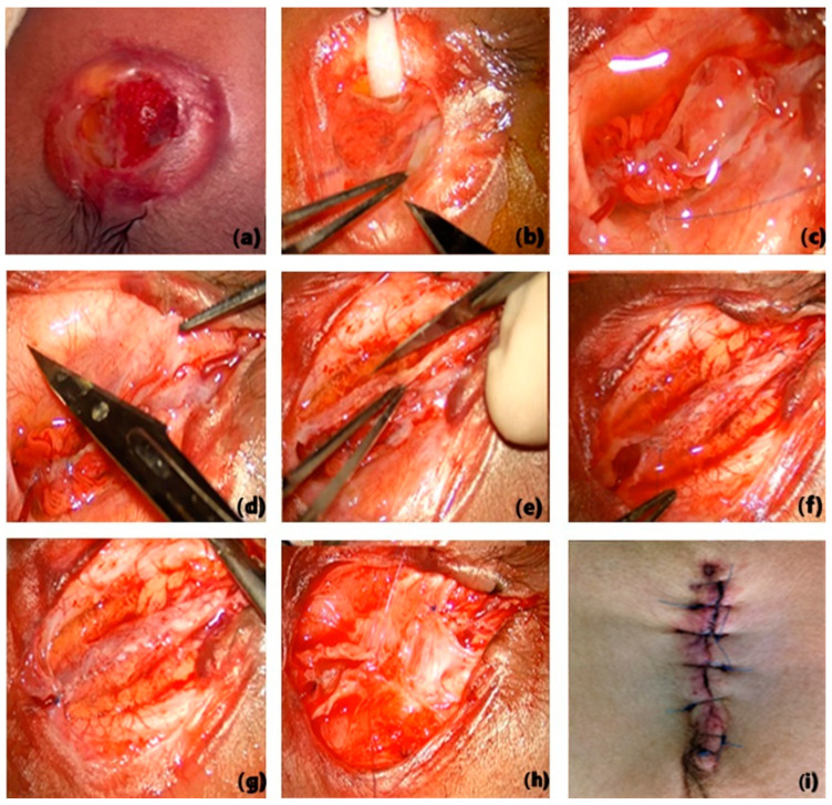
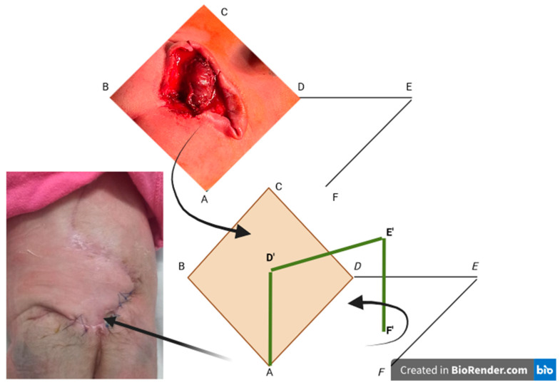
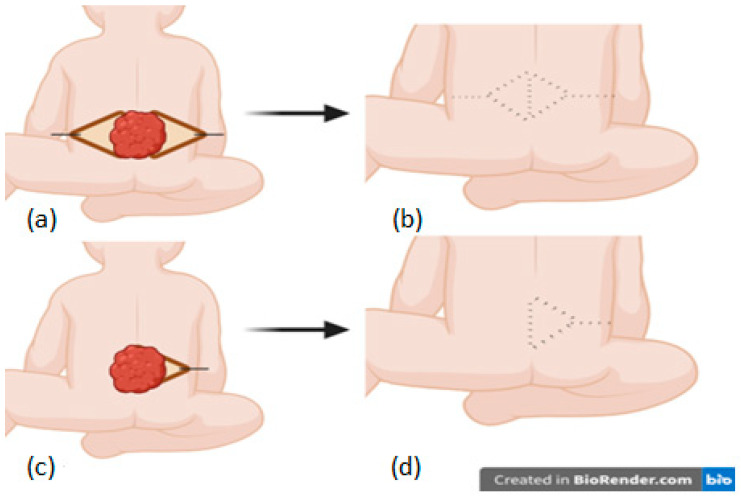
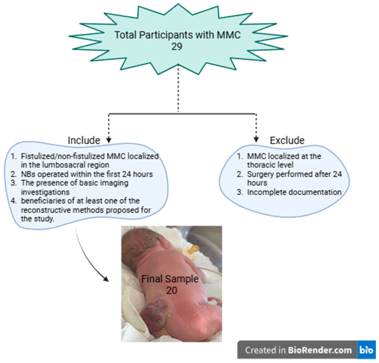
(1) Background: Neural tube defects are a prevalent cause of congenital malformations, myelomeningocele (MMC) being the most severe form. This study evaluates the clinical outcome and postoperative-associated complications following MMC surgical closures, focusing on the following three techniques: direct suture (DS); VY advancement flap (VYF); and Limberg flap (LF). (2) Methods: A retrospective observational study was conducted from March 2015 to February 2023, and the inclusion criteria were newborns who underwent lumbosacral MMC within 24 h of birth. (3) Results: Out of 20 cases, 45% underwent DS closure; 25% underwent VY-F closure; 15% underwent LF closure, and 15% (n = 3) underwent combined flap closure. A significant statistical correlation was observed between intracranial hypertension (IH), the need for external ventricular drainage (EVD), and DS closure type. In the DS group, 60% of patients required EVD (p = 0.041), and 90% had IH (p = 0.027). CSF fistula was present in 40% of LF cases and 30% of DS cases, while wound dehiscence was observed in 60% of LF cases and 30% of DS cases. (4) Conclusions: Our study demonstrated that DS was linked to higher rates of complications. The VY-F is the safest method for closing MMC defects.
Keywords: Limberg flap; VY flap; direct suture; dysraphism; myelomeningocele.
Conflict of interest statement
The authors declare no conflicts of interest.
Figures
References
-
- Paslaru F.G., Panaitescu A.M., Iancu G., Veduta A., Gica N., Paslaru A.C., Gheorghiu A., Peltecu G., Gorgan R.M. Myelomeningocele Surgery over the 10 Years Following the MOMS Trial: A Systematic Review of Outcomes in Prenatal versus Postnatal Surgical Repair. Medicina. 2021;57:707. doi: 10.3390/medicina57070707. - DOI - PMC - PubMed
Grants and funding
LinkOut - more resources
Full Text Sources
