

Assessment of Interrater Reliability and Accuracy of Cerebral Aneurysm Morphometry Using 3D Virtual Reality, 2D Digital Subtraction Angiography, and 3D Reconstruction: A Randomized Comparative Study
- PMID: 39451982
- PMCID: PMC11506597
- DOI: 10.3390/brainsci14100968
Assessment of Interrater Reliability and Accuracy of Cerebral Aneurysm Morphometry Using 3D Virtual Reality, 2D Digital Subtraction Angiography, and 3D Reconstruction: A Randomized Comparative Study
Abstract
Background/objectives: Detailed morphometric analysis of an aneurysm and the related vascular bifurcation are critical factors when determining rupture risk and planning treatment for unruptured intracranial aneurysms (UIAs). The standard visualization of digital subtraction angiography (DSA) and its 3D reconstruction on a 2D monitor provide precise measurements but are subject to variability based on the rater. Visualization using virtual (VR) and augmented reality platforms can overcome those limitations. It is, however, unclear whether accurate measurements of the aneurysm and adjacent arterial branches can be obtained on VR models. This study aimed to assess interrater reliability and compare measurements between 3D VR, standard 2D DSA, and 3D DSA reconstructions, evaluating the reliability and accuracy of 3D VR as a measurement tool.
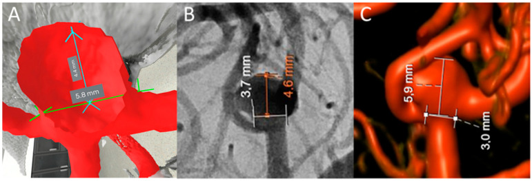
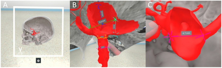
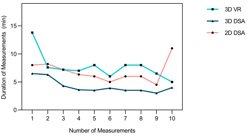
Methods: A pool of five neurosurgeons performed three individual analyses on each of the ten UIA cases, measuring them in completely immersed 3D VR and the standard on-screen format (2D DSA and 3D reconstruction). This resulted in three independent measurements per modality for each case. Interrater reliability of measurements and morphology characterization, comparative differences, measurement duration, and VR user experience were assessed.
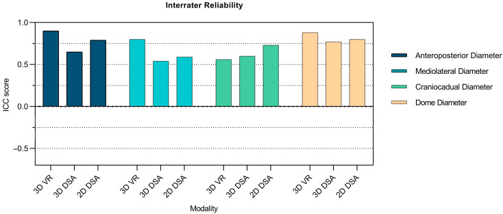
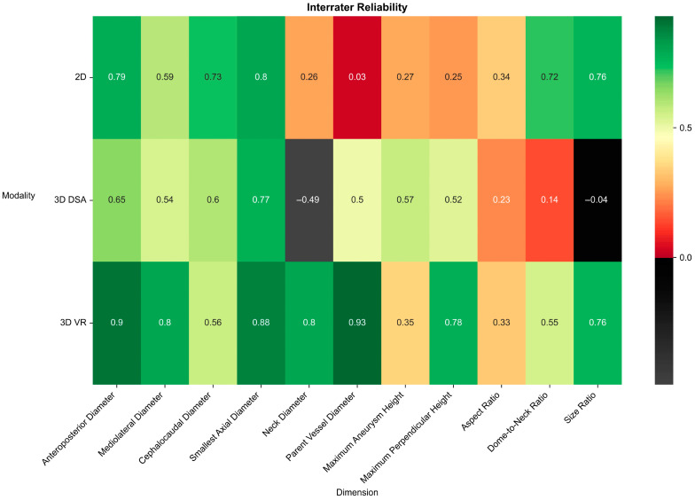
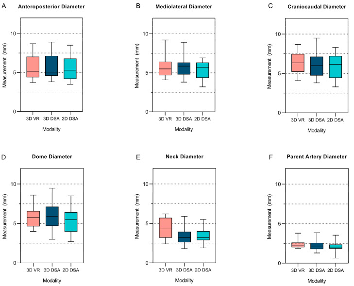
Results: Interrater reliability for 3D VR measurements was significantly higher than for 3D DSA measurements (3D VR mean intraclass correlation coefficient [ICC]: 0.69 ± 0.22 vs. 3D DSA mean ICC: 0.36 ± 0.37, p = 0.042). No significant difference was observed between 3D VR and 2D DSA (3D VR mean ICC: 0.69 ± 0.22 vs. 2D DSA mean ICC: 0.43 ± 0.31, p = 0.12). A linear mixed-effects model showed no effect of 3D VR and 3D DSA (95% CI = -0.26-0.28, p = 0.96) or 3D VR and 2D DSA (95% CI = -0.02-0.53, p = 0.066) on absolute measurements of the aneurysm in the anteroposterior, mediolateral, and craniocaudal dimensions.
Conclusions: 3D VR technology allows for reproducible, accurate, and reliable measurements comparable to measurements performed on a 2D screen. It may also potentially improve precision for measurements of non-planar aneurysm dimensions.
Keywords: augmented reality; cerebrovascular neurosurgery; intracranial aneurysm; measurement; morphometry; surgical planning; virtual reality.
Conflict of interest statement
The authors declare no conflicts of interest.
Figures

References
LinkOut - more resources
Full Text Sources
