

Estimated Disease Progression Trajectory of White Matter Disruption in Unilateral Temporal Lobe Epilepsy: A Data-Driven Machine Learning Approach
- PMID: 39452006
- PMCID: PMC11506697
- DOI: 10.3390/brainsci14100992
Estimated Disease Progression Trajectory of White Matter Disruption in Unilateral Temporal Lobe Epilepsy: A Data-Driven Machine Learning Approach
Abstract
Background/objectives: Although the involvement of progressive brain alterations in epilepsy was recently suggested, individual patients' trajectories of white matter (WM) disruption are not known.
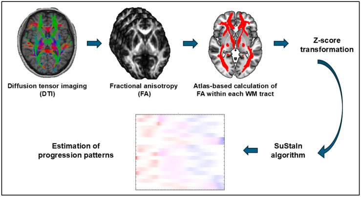
Methods: We investigated the disease progression patterns of WM damage and its associations with clinical metrics. We examined the cross-sectional diffusion tensor imaging (DTI) data of 155 patients with unilateral temporal lobe epilepsy (TLE) and 270 age/gender-matched healthy controls, and we then calculated the average fractional anisotropy (FA) values within 20 WM tracts of the whole brain. We used the Subtype and Stage Inference (SuStaIn) program to detect the progression trajectory of FA changes and investigated its association with clinical parameters including onset age, disease duration, drug-responsiveness, and the number of anti-seizure medications (ASMs).
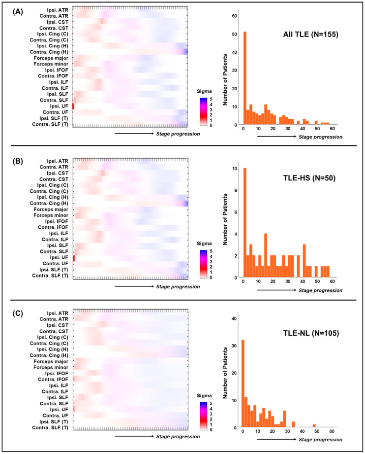
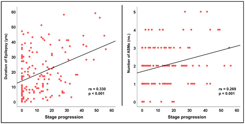
Results: The SuStaIn algorithm identified a single subtype model in which the initial damage occurs in the ipsilateral uncinate fasciculus (UF), followed by damage in the forceps, superior longitudinal fasciculus (SLF), and anterior thalamic radiation (ATR). This pattern was replicated when analyzing TLE with hippocampal sclerosis (n = 50) and TLE with no lesions (n = 105) separately. Further-progressed stages were associated with longer disease duration (p < 0.001) and a greater number of ASMs (p = 0.001).
Conclusions: the disease progression model based on WM tracts may be useful as a novel individual-level biomarker.
Keywords: diffusion tensor imaging; machine learning; temporal lobe epilepsy; white matter.
Conflict of interest statement
The authors declare no conflicts of interest.
Figures
Similar articles
-
White matter abnormalities associate with type and localization of focal epileptogenic lesions.Epilepsia. 2015 Jan;56(1):125-32. doi: 10.1111/epi.12871. Epub 2014 Dec 26. Epilepsia. 2015. PMID: 25545559
-
Spatial patterns of gray and white matter compromise relate to age of seizure onset in temporal lobe epilepsy.Neuroimage Clin. 2023;39:103473. doi: 10.1016/j.nicl.2023.103473. Epub 2023 Jul 8. Neuroimage Clin. 2023. PMID: 37531834 Free PMC article.
-
MRI-Derived Modeling of Disease Progression Patterns in Patients With Temporal Lobe Epilepsy.Neurology. 2024 Aug 13;103(3):e209524. doi: 10.1212/WNL.0000000000209524. Epub 2024 Jul 9. Neurology. 2024. PMID: 38981074
-
Association of white matter diffusion characteristics and cognitive deficits in temporal lobe epilepsy.Epilepsy Behav. 2018 Feb;79:138-145. doi: 10.1016/j.yebeh.2017.11.040. Epub 2018 Jan 4. Epilepsy Behav. 2018. PMID: 29287217
-
The role of diffusion tensor imaging and fractional anisotropy in the evaluation of patients with idiopathic normal pressure hydrocephalus: a literature review.Neurosurg Focus. 2016 Sep;41(3):E12. doi: 10.3171/2016.6.FOCUS16192. Neurosurg Focus. 2016. PMID: 27581308 Review.
References
Grants and funding
LinkOut - more resources
Full Text Sources
Miscellaneous
