

Contrast Sensitivity Is Impaired in Suspected Primary Open-Angle Glaucoma Patients
- PMID: 39452007
- PMCID: PMC11505721
- DOI: 10.3390/brainsci14100993
Contrast Sensitivity Is Impaired in Suspected Primary Open-Angle Glaucoma Patients
Abstract
Purpose: To assess spatial contrast sensitivity (CS) in suspected primary open-angle glaucoma (POAG) patients.
Methods: CS was measured using sinusoidal gratings of 4 cycles/degree. First, foveal and peripheral CS were assessed in 34 suspected POAG patients and compared with 71 and 28 age-matched healthy individuals for foveal and peripheral conditions, respectively. Second, foveal CS was assessed in 34 early POAG patients age-matched with suspected POAG patients. Analyses were performed considering two age ranges: Under and Over 50 y.o. Correlations were evaluated between CS and clinical parameters. Diagnostic accuracy was also analyzed.
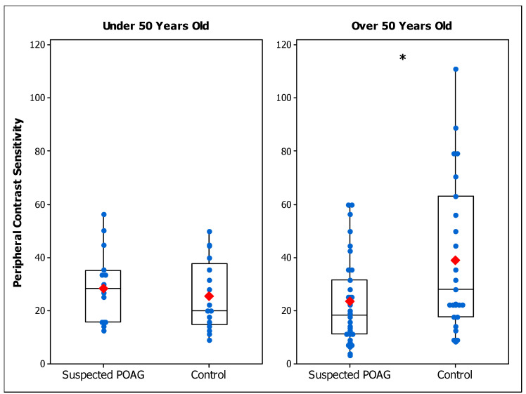
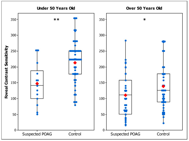
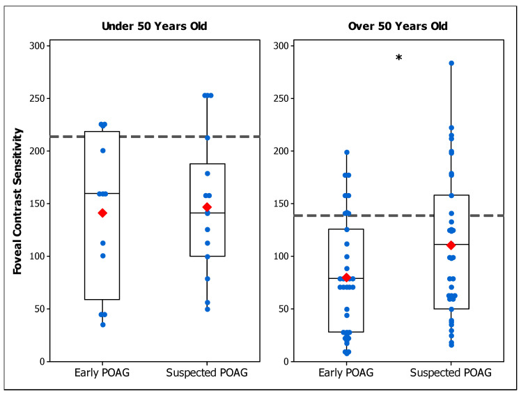
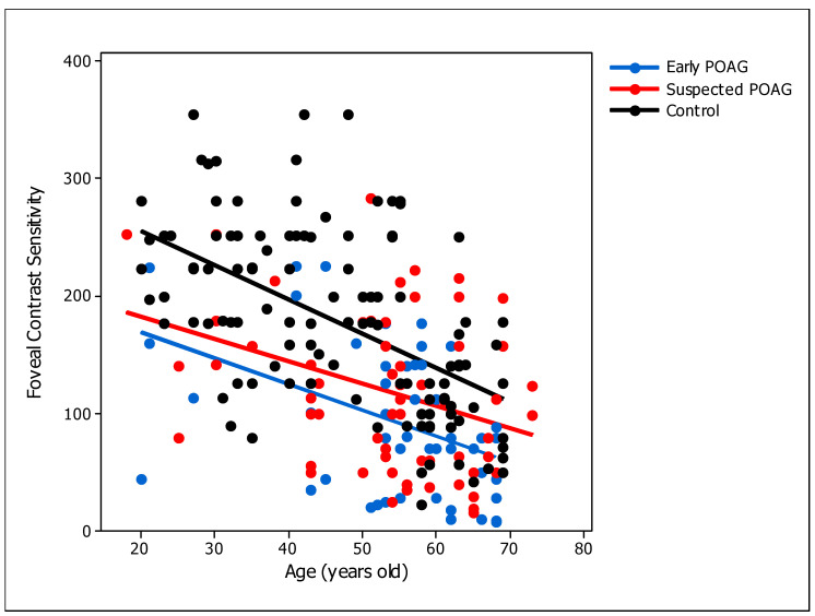
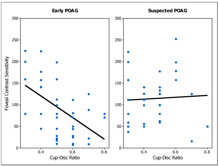
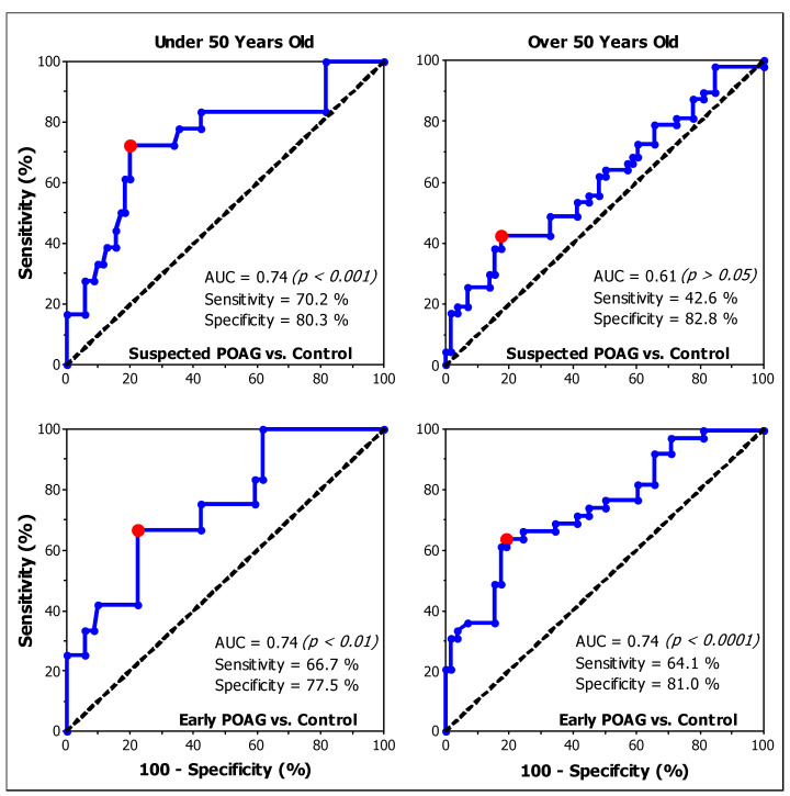
Results: Peripheral CS was lower in older suspected POAG patients (23.4 ± 16.1) than the control group (39.1 ± 28.2) (p = 0.040). Foveal CS was reduced in suspected POAG participants (Under 50: 146.8 ± 63.3; p = 0.004. Over 50: 110.5 ± 65.0; p = 0.044) and in early POAG patients (Under 50: 141.2 ± 72.6; p = 0.002. Over 50: 80.2 ± 54.5 p < 0.001), both compared to the control group (Under 50: 213.5 ± 66.2. Over 50: 138.6 ± 71.7). CS was lower in early POAG than in POAG suspected in older patients (p = 0.042). Foveal CS was correlated with age (Early: p = 0.001. Suspect: p = 0.002) and with the cup-disc ratio only in early POAG patients (p < 0.001). Foveal CS had fair (AUC = 0.74) diagnostic accuracy for early POAG patients.
Conclusions: CS in suspected POAG patients is lower than in healthy individuals. Our findings evidence the spatial vision loss before the onset of POAG.
Keywords: age; contrast sensitivity; early detection; functional assessment; glaucoma.
Conflict of interest statement
The authors declare no conflicts of interest.
Figures
Similar articles
-
Value of disc topographic measurements for the discrimination of glaucoma suspects from early primary open angle glaucoma cases: A thin discriminative line.Photodiagnosis Photodyn Ther. 2023 Sep;43:103746. doi: 10.1016/j.pdpdt.2023.103746. Epub 2023 Aug 17. Photodiagnosis Photodyn Ther. 2023. PMID: 37595654
-
Comparing glaucomatous optic neuropathy in primary open angle and chronic primary angle closure glaucoma eyes by optical coherence tomography.Ophthalmic Physiol Opt. 2005 Sep;25(5):408-15. doi: 10.1111/j.1475-1313.2005.00304.x. Ophthalmic Physiol Opt. 2005. PMID: 16101946
-
Routine eye examinations for persons 20-64 years of age: an evidence-based analysis.Ont Health Technol Assess Ser. 2006;6(15):1-81. Epub 2006 Jul 1. Ont Health Technol Assess Ser. 2006. PMID: 23074485 Free PMC article.
-
Differences in Optic Disc Characteristics of Primary Congenital Glaucoma, Juvenile, and Adult Onset Open Angle Glaucoma Patients.J Glaucoma. 2016 Mar;25(3):239-43. doi: 10.1097/IJG.0000000000000154. J Glaucoma. 2016. PMID: 25265002
-
Pattern Electroretinography Changes in Patients with Established or Suspected Primary Open Angle Glaucoma.J Curr Glaucoma Pract. 2013 May-Aug;7(2):39-42. doi: 10.5005/jp-journals-10008-1135. Epub 2013 May 9. J Curr Glaucoma Pract. 2013. PMID: 26997780 Free PMC article. Review.
Cited by
-
The mesopic negative response (MeNR): a novel approach to assess retinal ganglion cell function within the rod pathway.Doc Ophthalmol. 2025 Jul 9. doi: 10.1007/s10633-025-10040-3. Online ahead of print. Doc Ophthalmol. 2025. PMID: 40634831
References
Grants and funding
LinkOut - more resources
Full Text Sources
