

A Dual Role for the Dorsolateral Prefrontal Cortex (DLPFC) in Auditory Deviance Detection
- PMID: 39452008
- PMCID: PMC11505713
- DOI: 10.3390/brainsci14100994
A Dual Role for the Dorsolateral Prefrontal Cortex (DLPFC) in Auditory Deviance Detection
Abstract
Background: In the oddball paradigm, the dorsolateral prefrontal cortex (DLPFC) is often associated with active cognitive responses, such as maintaining information in working memory or adapting response strategies. While some evidence points to the DLPFC's role in passive auditory deviance perception, a detailed understanding of the spatiotemporal neurodynamics involved remains unclear.
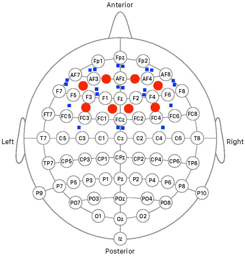
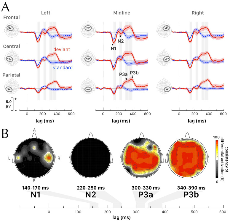
Methods: In this study, event-related optical signals (EROS) and event-related potentials (ERPs) were simultaneously recorded for the first time over the prefrontal cortex using a 64-channel electroencephalography (EEG) system, during passive auditory deviance perception in 12 right-handed young adults (7 women and 5 men). In this oddball paradigm, deviant stimuli (a 1500 Hz pure tone) elicited a negative shift in the N1 ERP component, related to mismatch negativity (MMN), and a significant positive deflection associated with the P300, compared to standard stimuli (a 1000 Hz tone).
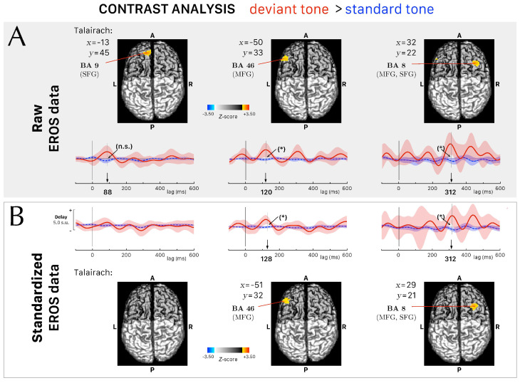
Results: We hypothesize that the DLPFC not only participates in active tasks but also plays a critical role in processing deviant stimuli in passive conditions, shifting from pre-attentive to attentive processing. We detected enhanced neural activity in the left middle frontal gyrus (MFG), at the same timing of the MMN component, followed by later activation at the timing of the P3a ERP component in the right MFG.
Conclusions: Understanding these dynamics will provide deeper insights into the DLPFC's role in evaluating the novelty or unexpectedness of the deviant stimulus, updating its cognitive value, and adjusting future predictions accordingly. However, the small number of subjects could limit the generalizability of the observations, in particular with respect to the effect of handedness, and additional studies with larger and more diverse samples are necessary to validate our conclusions.
Keywords: BA 46; BA 8; P300; frequency-domain fNIRS; mismatch negativity; oddball paradigm; optical imaging.
Conflict of interest statement
The authors declare no conflicts of interest.
Figures
Similar articles
-
The spatio-temporal dynamics of deviance and target detection in the passive and active auditory oddball paradigm: a sLORETA study.BMC Neurosci. 2018 Apr 19;19(1):25. doi: 10.1186/s12868-018-0422-3. BMC Neurosci. 2018. PMID: 29673322 Free PMC article.
-
Preattentive cortical-evoked responses to pure tones, harmonic tones, and speech: influence of music training.Ear Hear. 2009 Aug;30(4):432-46. doi: 10.1097/AUD.0b013e3181a61bf2. Ear Hear. 2009. PMID: 19494778
-
Auditory pre-attentive processing of Chinese tones.Chin Med J (Engl). 2008 Dec 5;121(23):2429-33. Chin Med J (Engl). 2008. PMID: 19102963 Clinical Trial.
-
Pre-attentive Mismatch Response and Involuntary Attention Switching to a Deviance in an Earlier-Than-Usual Auditory Stimulus: An ERP Study.Front Hum Neurosci. 2019 Mar 6;13:58. doi: 10.3389/fnhum.2019.00058. eCollection 2019. Front Hum Neurosci. 2019. PMID: 30894807 Free PMC article.
-
Detection of stimulus deviance within primate primary auditory cortex: intracortical mechanisms of mismatch negativity (MMN) generation.Brain Res. 1994 Dec 26;667(2):192-200. doi: 10.1016/0006-8993(94)91496-6. Brain Res. 1994. PMID: 7697356
Cited by
-
Brain Activation During Motor Preparation and Execution in Patients with Mild Cognitive Impairment: An fNIRS Study.Brain Sci. 2025 Mar 23;15(4):333. doi: 10.3390/brainsci15040333. Brain Sci. 2025. PMID: 40338241 Free PMC article.
References
-
- Maturana H.R., Varela F.J. Autopoiesis and Cognition: The Realization of the Living. Volume 42 D. Reidel Publishing Company; Dordrecht, Holland: 1980.
Grants and funding
LinkOut - more resources
Full Text Sources
Miscellaneous
