Review
. 2024 Oct 16;14(10):1022.
doi: 10.3390/brainsci14101022.

Strategic Integration: A Cross-Disciplinary Review of the fNIRS-EEG Dual-Modality Imaging System for Delivering Multimodal Neuroimaging to Applications

Affiliations
Review

Strategic Integration: A Cross-Disciplinary Review of the fNIRS-EEG Dual-Modality Imaging System for Delivering Multimodal Neuroimaging to Applications

Jiafa Chen et al. Brain Sci. .

Abstract

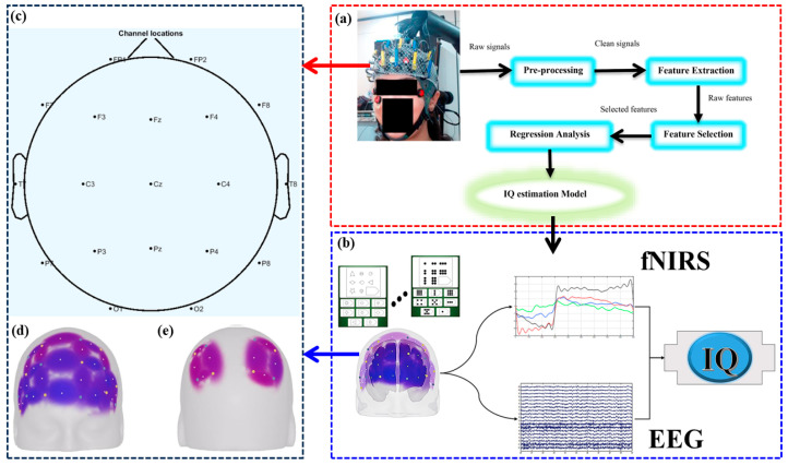
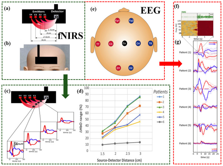
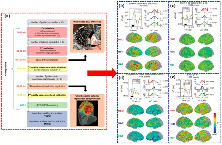
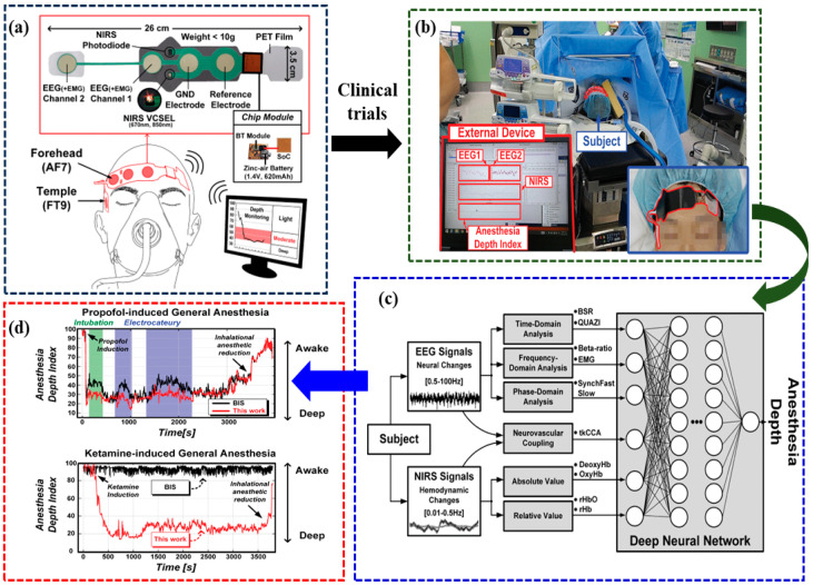
Background: Recent years have seen a surge of interest in dual-modality imaging systems that integrate functional near-infrared spectroscopy (fNIRS) and electroencephalography (EEG) to probe brain function. This review aims to explore the advancements and clinical applications of this technology, emphasizing the synergistic integration of fNIRS and EEG. Methods: The review begins with a detailed examination of the fundamental principles and distinctive features of fNIRS and EEG techniques. It includes critical technical specifications, data-processing methodologies, and analysis techniques, alongside an exhaustive evaluation of 30 seminal studies that highlight the strengths and weaknesses of the fNIRS-EEG bimodal system. Results: The paper presents multiple case studies across various clinical domains-such as attention-deficit hyperactivity disorder, infantile spasms, depth of anesthesia, intelligence quotient estimation, and epilepsy-demonstrating the fNIRS-EEG system's potential in uncovering disease mechanisms, evaluating treatment efficacy, and providing precise diagnostic options. Noteworthy research findings and pivotal breakthroughs further reinforce the developmental trajectory of this interdisciplinary field. Conclusions: The review addresses challenges and anticipates future directions for the fNIRS-EEG dual-modal imaging system, including improvements in hardware and software, enhanced system performance, cost reduction, real-time monitoring capabilities, and broader clinical applications. It offers researchers a comprehensive understanding of the field, highlighting the potential applications of fNIRS-EEG systems in neuroscience and clinical medicine.

Keywords: EEG; clinical application; dual-mode monitoring; fNIRS.


Conflict of interest statement

The authors have no relevant financial or non-financial interests to disclose.

Figures

Figure 1
Figure 1
Comparison of the resolutions of commonly used functional brain-imaging techniques.
Figure 2
Figure 2
fNIRS-EEG dual-modality imaging system fusion method for assessing brain functions.
Figure 3
Figure 3
Accompanying symptoms of ADHD in children and adolescents.
Figure 4
Figure 4
Visualization of the fNIRS-EEG dual-modality imaging system for diagnosing children with ADHD [29]. (a) EEG electrodes were placed on the Fz (frontal), Pz (parietal), Oz (occipital) and Cz (central) locations according to the international 10–20 system. A band with optical fiber probes was placed on the forehead for fNIRS data acqusition. Data acquisition system has been shown on a control subject. (b) Spatial profiles of the fNIRS channels and the ROIs locations. (c) The source-detector and 16 optode (channel) measurement locations registered on fNIR probe. (d) The flowchart of the study (the signals belong to a random control and ADHD subject). (e) ROC Curve for MLP (AUC = 0.92). (f) ROC Curve for SVM (AUC = 0.859). (g) ROC Curve for NB (AUC = 0.937).
Figure 5
Figure 5
Visualization of the fNIRS-EEG dual-modality imaging system for studying IQ estimation [49]. (a) The overall scheme of the IQ estimation procedure. (b) IQ Experiment Flowchart. (c) the location of EEG electrodes. (d) fNIRS Optodes placement (e) channel configuration for problem solving task.
Figure 6
Figure 6
Visualization of the fNIRS-EEG dual-modality imaging system for studying infantile spasms [57]. (a) fNIRS: a patch composed of four pairs of optical fibers (each wavelength corresponds to one thread in each team), containing four transmitters and one detector. (b) fNIRS: a detector has been positioned in the middle of the forehead. (c) fNIRS: Hemodynamics observed over multiple distances via fNIRS spectroscopic technique graphs. (d) fNIRS: Normalized range values of [HbO] for the four source-detector distances in the period of −5 to 25 s vs source-detector distances (1.5, 2, 2.5, 3 cm) for the 6 patients. (e) EEG: layout of nine electrodes (10–20 system configurations, with a frontal reference). (f) A time-frequency response (TFR) of the deltoid EMG determined the onset of each infantile spasms (T0). infantile spasms onset was always characterized by a sudden increase in the deltoid EMG power of all frequency bands between 0 and 100 Hz. (g) A two-phase hemodynamic change started with the onset of EMG activation (as determined in a time-frequency analysis).
Figure 7
Figure 7
Visualization of the fNIRS-EEG dual-modality imaging system for the study of epilepsy [67]. (a) Sketch plot of the patient evaluation, continuous encephalography and fNIRS data acquisition, and data analysis procedures (b) Hemodynamic changes associated with nonconvulsive seizures. (c) Hemodynamic changes associated with BS bursts. (d) Hemodynamic changes associated with burst suppression suppressions. (e) Hemodynamic changes associated with periodic discharges.
Figure 8
Figure 8
Visualization of the fNIRS-EEG dual-modality imaging system for studying depth of anesthesia [69]. (a) Proposed multimodal anesthesia depth monitoring system. (b) Clinical trial environment. (c) Flowchart of the deep-learning algorithm. (d) Clinical results for propofol-induced general anes-thesia and ketamine-induced general anesthesia.

Similar articles

Cited by

References

    1. Hong K.S., Khan M.J., Hong M.J. Feature Extraction and Classification Methods for Hybrid fNIRS-EEG Brain-Computer Interfaces. Front. Hum. Neurosci. 2018;12:25. doi: 10.3389/fnhum.2018.00246. - DOI - PMC - PubMed
    1. Li R.H., Yang D.L., Fang F., Hong K.S., Reiss A.L., Zhang Y.C. Concurrent fNIRS and EEG for Brain Function Investigation: A Systematic, Methodology-Focused Review. Sensors. 2022;22:5865. doi: 10.3390/s22155865. - DOI - PMC - PubMed
    1. Liu Z.M., Shore J., Wang M., Yuan F.P., Buss A., Zhao X.P. A systematic review on hybrid EEG/fNIRS in brain-computer interface. Biomed. Signal Process. Control. 2021;68:8. doi: 10.1016/j.bspc.2021.102595. - DOI
    1. Kassab A., Le Lan J., Tremblay J., Vannasing P., Dehbozorgi M., Pouliot P., Gallagher A., Lesage F., Sawan M., Nguyen D.K. Multichannel wearable fNIRS-EEG system for long-term clinical monitoring. Hum. Brain Mapp. 2018;39:7–23. doi: 10.1002/hbm.23849. - DOI - PMC - PubMed
    1. Shin S.S., Bales J.W., Edward Dixon C., Hwang M. Structural imaging of mild traumatic brain injury may not be enough: Overview of functional and metabolic imaging of mild traumatic brain injury. Brain Imaging Behav. 2017;11:591–610. doi: 10.1007/s11682-017-9684-0. - DOI - PubMed

LinkOut - more resources