Review
. 2024 Sep 28;11(10):300.
doi: 10.3390/jcdd11100300.

Storytelling of Hypertrophic Cardiomyopathy Discovery

Affiliations
Review

Storytelling of Hypertrophic Cardiomyopathy Discovery

Gaetano Thiene et al. J Cardiovasc Dev Dis. .

Abstract

The discovery of hypertrophic cardiomyopathy (HCM) dates back to 1958, when the pathologist Donald Teare of the St. George's Hospital in London performed autopsies in eight cases with asymmetric hypertrophy of the ventricular septum and bizarre disorganization (disarray) at histology, first interpreted as hamartoma. Seven had died suddenly. The cardiac specimens were cut along the long axis, similar to the 2D echo. In the same year, at the National Institute of Health U.S.A., Eugene Braunwald, a hemodynamist, and Andrew Glenn Morrow, a cardiac surgeon, clinically faced a patient with an apparently similar morbid entity, with a systolic murmur and subaortic valve gradient. "Discrete" subaortic stenosis was postulated. However, at surgery, Dr. Morrow observed only hypertrophy and performed myectomy to relieve the obstruction. This first Braunwald-Morrow patient underwent a successful cardiac transplant later at the disease end stage. The same Dr. Morrow was found to be affected by the familial HCM and died suddenly in 1992. The term "functional subaortic stenosis" was used in 1959 and "idiopathic hypertrophic subaortic stenosis" in 1960. Years before, in 1957, Lord Brock, a cardiac surgeon at the Guy's Hospital in London, during alleged aortic valve surgery in extracorporeal circulation, did not find any valvular or discrete subaortic stenoses. In 1980, John F. Goodwin of the Westminster Hospital in London, the head of an international WHO committee, put forward the first classification of heart muscle diseases, introducing the term cardiomyopathy (dilated, hypertrophic, and endomyocardial restrictive). In 1995, the WHO classification was revisited, with the addition of two new entities, namely arrhythmogenic and purely myocardial restrictive, the latter a paradox of a small heart accounting for severe congestive heart failure by ventricular diastolic impairment. A familial occurrence was noticed earlier in HCM and published by Teare and Goodwin in 1960. In 1989-1990, the same family underwent molecular genetics investigation by the Seidman team in Boston, and a missense mutation of the β-cardiac myosin heavy chain in chromosome 14 was found. Thus, 21 years elapsed from HCM gross discovery to molecular discoveries. The same original family was the source of both the gross and genetic explanations of HCM, which is now named sarcomere disease. Restrictive cardiomyopathy, characterized grossly without hypertrophy and histologically by myocardial disarray, was found to also have a sarcomeric genetic mutation, labeled "HCM without hypertrophy". Sarcomere missense mutations have also been reported in dilated cardiomyopathy (DCM) and non-compaction cardiomyopathy. Moreover, sarcomeric gene defects have been detected in some DNA non-coding regions of HCM patients. The same mutation in the family may express different phenotypes (HCM, DCM, and RCM). Large ischemic scars have been reported by pathologists and are nowadays easily detectable in vivo by cardiac magnetic resonance with gadolinium. The ischemic arrhythmic substrate enhances the risk of sudden death.

Keywords: cardiomyopathies; history of medicine; hypertrophic cardiomyopathy; pathology; restrictive cardiomyopathy; sudden cardiac death; transplantation.


Conflict of interest statement

The authors declare no conflicts of interest.

Figures

Figure 1
Figure 1
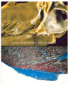
(a) The original picture of a cardiac specimen from the Donald Teare paper [1]. Coincidentally, the cut was performed along the long axis of the left ventricle, similar to the current 2D echo tomographic section, depicting the septal asymmetric hypertrophy (arrow). (b) Myocardial disarray under a light microscope, mimicking a hamartoma. Azan-Mallory stain, low magnification.
Figure 2
Figure 2
Picture of the young Eugene Braunwald in the 1960s at the National Institutes of Health (NIH), chief of hemodynamics.
Figure 3
Figure 3
(a) Mr. Brady’s continuous pullback pressure tracing, recorded as the catheter was withdrawn from the LV cavity through the outflow tract across the aortic valve and into the aorta. Reproduced with permission from [2]. (b) Andrew Glenn Morrow’s picture, chief of Cardiac Surgery at the NIH in the 1960s. He wrote the paper reporting the first patient with HCM and subaortic gradient [3].
Figure 4
Figure 4
The surgical myectomy technique, invented by Morrow.
Figure 5
Figure 5
A recent picture of Mr. Brady, the first patient in the USA diagnosed as affected by idiopathic hypertrophic subaortic stenosis (with Dr. Bonow (left), Mrs. Salberg (middle), and Dr. Maron (right)) [6].
Figure 6
Figure 6
John F. Goodwin, Chief of Cardiology at the Hammersmith Hospital in London, who in 1980 published the first WHO classification of primary heart muscle diseases, introducing the term cardiomyopathy [7].
Figure 7
Figure 7
The three cardiomyopathies of the 1980 WHO classification: dilated, hypertrophic, and restrictive, the latter referring to Loeffer eosinophilic endomyocardial disease.
Figure 8
Figure 8
Morrow Family Pedigree. Family genealogic tree of Dr. Morrow, who was affected by hypertrophic cardiomyopathy and died suddenly in 1982. Squares indicate men; circles indicate women; the arrow indicates the proband (Dr. Morrow, I.1.); solid symbols indicate hypertrophic cardiomyopathy; and the slash indicates deceased.
Figure 9
Figure 9
Familial genealogic tree of a young patient who died suddenly in the original series Teare published in 1960 [11].
Figure 10
Figure 10
Sarcomeric proteins, carried by missense mutations of causative genes in HCM (from [13]), with permission.
Figure 11
Figure 11
Image of 2D echocardiography, the revolutionary non-invasive diagnostic technique for an easy visualization of asymmetric hypertrophy. (a) Schematic long-axis tomographic view at 2D cardiac echo. (b) The equivalent in a normal heart. (c) The 2D echo in hypertrophic cardiomyopathy. (d) Cardiac specimen of (c), with septal asymmetric hypertrophy. A = aorta; AA = ascending aorta; AV = aortic valve; LA = left atrium; LV = left ventricle; PW = posterior wall; RV = right ventricle; and VS = ventricular septum.
Figure 12
Figure 12
Subaortic endocardial fibrotic plaque (arrow) due to friction between the mitral anterior leaflet and the asymmetric septal hypertrophy.
Figure 13
Figure 13
Mechanism accounting for mitral regurgitation and the dilatation of the left atrium in hypertrophic cardiomyopathy. LA = left atrium; LV = left ventricle; and MV = mitral valve.
Figure 14
Figure 14
Thrombosis in the left atrial appendage (arrow) of a patient with hypertrophic cardiomyopathy, dilated left atrium, and atrial fibrillation who underwent cardiac transplantation. LA = left atrium; LV = left ventricle; RA = right atrium; and RV = right ventricle.
Figure 15
Figure 15
A figure from Teare’s original paper [1] representing a cardiac specimen with a dilated left atrium. LA = left atrium; MV = mitral valve.
Figure 16
Figure 16
Infective endocarditis (IE), developed on subaortic septal plaque, in a patient with hypertrophic cardiomyopathy. The vegetations conveyed the infection to the anterior leaflet mitral valve through a “kissing” mechanism, with perforation (arrow) and aneurysm. The infection also involved the aortic valve.
Figure 17
Figure 17
Infarct-like scars of the ventricular septum and free wall of the left ventricle in end-stage hypertrophic cardiomyopathy.
Figure 18
Figure 18
Acute ischemic myocardial damage in hypertrophic cardiomyopathy at various histologic stages. Hematoxylin and eosin stains at high (b) and low (a,c,d) magnifications.
Figure 19
Figure 19
(a,b) Massive infarction of the ventricular septum. Note the huge asymmetric hypertrophy (arrows) and (c) deep, long intramural course of the anterior descending coronary artery (arrow).
Figure 20
Figure 20
This is the original illustration of the publication of the intramural course of the anterior descending coronary artery (ADCA) as an isolated cause of sudden death. However, it is quite evident that it is a case of hypertrophic cardiomyopathy with anteroseptal asymmetric hypertrophy and intramural course (arrow) of ADCA. Permission was obtained from [27].
Figure 21
Figure 21
Restrictive cardiomyopathy with normal ventricles, dilated atria (a), and ventricular myocardial disarray at histology (b). Azan-Mallory stain at high magnification. LA = left atrium; LV = left ventricle; RA = right atrium; RV = right ventricle.
Figure 22
Figure 22
The paradox of a small heart in restrictive cardiomyopathy (on the right) requiring transplantation, compared to the “cor bovinum” of dilated cardiomyopathy (on the left). A genetic mutation of troponin I (low) was found [14]. Tm= tropomyosin; TnC = troponin C; TnI = troponin I; TnT = troponin T.

References

    1. Teare D. Asymmetrical hypertrophy of the heart in young adults. Br. Heart J. 1958;20:1–8. doi: 10.1136/hrt.20.1.1. - DOI - PMC - PubMed
    1. Braunwald E. Reflections on Hypertrophic Cardiomyopathy. Eur. Heart J. 2021;42:2969–2970. doi: 10.1093/eurheartj/ehab337. - DOI - PubMed
    1. Morrow A.G., Braunwald E. Functional aortic stenosis; a malformation characterized by resistance to left ventricular outflow without anatomic obstruction. Circulation. 1959;20:181–189. doi: 10.1161/01.CIR.20.2.181. - DOI - PubMed
    1. Aygen M.M., Hilbish T.F., Braunwald E., Morrow A.G., Cornell W.P. Idiopathic hypertrophic subaortic stenosis: Clinical, hemodynamic and angiographic manifestations. Am. J. Med. 1960;29:924–945.
    1. Brock R. Functional obstruction of the left ventricle; acquired aortic subvalvar stenosis. Guys Hosp. Rep. 1957;106:221–238. - PubMed

LinkOut - more resources