

The Role of Plain Radiography in Assessing Aborted Foetal Musculoskeletal Anomalies in Everyday Practice
- PMID: 39452405
- PMCID: PMC11508908
- DOI: 10.3390/jimaging10100242
The Role of Plain Radiography in Assessing Aborted Foetal Musculoskeletal Anomalies in Everyday Practice
Abstract
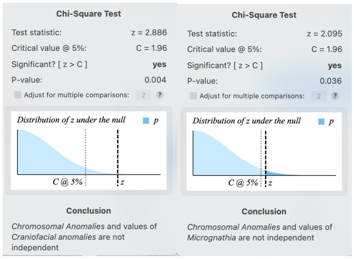
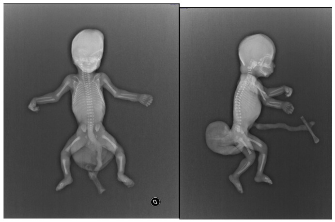
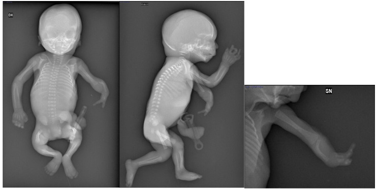
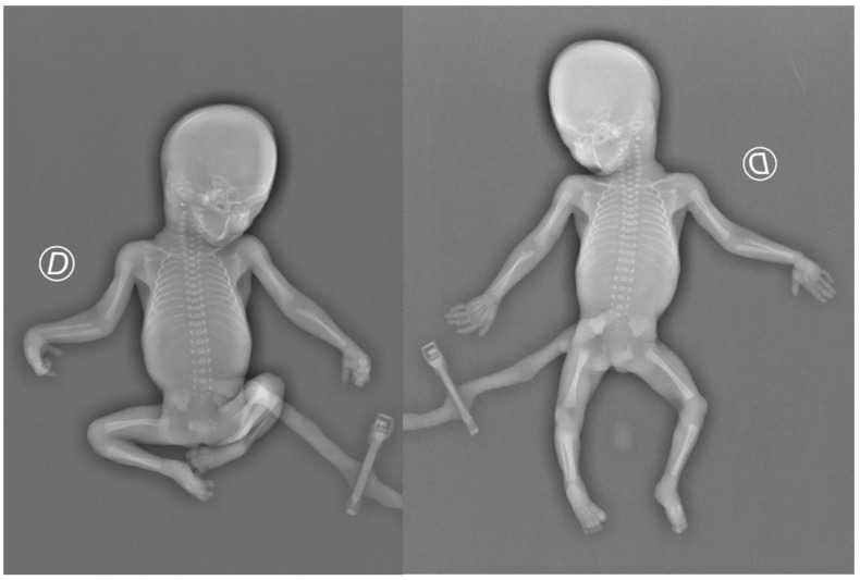
Conventional radiography is widely used for postmortem foetal imaging, but its role in diagnosing congenital anomalies is debated. This study aimed to assess the effectiveness of X-rays in detecting skeletal abnormalities and guiding genetic analysis and counselling. This is a retrospective analysis of all post-abortion diagnostic imaging studies conducted at a centre serving a population of over 300,000 inhabitants from 2008 to 2023. The data were analysed using descriptive statistics. X-rays of 81 aborted foetuses (total of 308 projections; mean: 3.8 projections/examination; SD: 1.79) were included. We detected 137 skeletal anomalies. In seven cases (12.7%), skeletal anomalies identified through radiology were missed by prenatal sonography. The autopsy confirmed radiological data in all cases except for two radiological false positives. Additionally, radiology failed to identify a case of syndactyly, which was revealed by anatomopathology. X-ray is crucial for accurately classifying skeletal abnormalities, determining the causes of spontaneous abortion, and guiding the request for genetic counselling. Formal training for both technicians and radiologists, as well as multidisciplinary teamwork, is necessary to perform X-ray examinations on aborted foetuses and interpret the results effectively.
Keywords: X-ray; diagnostic algorithms; projections; skeletal.
Conflict of interest statement
The authors declare no conflicts of interest.
Figures
References
-
- Odendaal H.J., Elliott A., Kinney H.C., Human M., Gaspar D., Petersen D., Randall B., Dempers J. Prenatal Alcohol and SIDS and Stillbirth (PASS) Network. Consent for autopsy research for unexpected death in early life. Obstet. Gynecol. 2011;117:167–171. doi: 10.1097/AOG.0b013e318200cb17. - DOI - PMC - PubMed
LinkOut - more resources
Full Text Sources
