Case Reports
. 2024 Sep 24;14(10):1019.
doi: 10.3390/jpm14101019.

Preoperative Embolization in the Management of Giant Thoracic Tumors: A Case Series

Affiliations
Case Reports

Preoperative Embolization in the Management of Giant Thoracic Tumors: A Case Series

Nicola Maria Lucarelli et al. J Pers Med. .

Abstract

Objectives: The aim of this paper is to describe our experience in the embolization of hypervascular giant thoracic tumors before surgical excision. Methods: A single-center retrospective review of five trans-arterial preoperative embolization procedures executed between October 2020 and July 2024. Patients' demographics, anatomical aspects, feasibility, technique, and outcomes were reviewed. Results: In all cases, accurate targeting and safe embolization was achieved, with satisfactory devascularization evaluated with post-procedural angiography and with minimal blood loss during subsequent surgical operation. Conclusions: In our experience, preoperative embolization of giant thoracic masses has been technically feasible, safe, and effective in reducing tumor vascularization, thus facilitating surgical treatment. This approach should be evaluated as an option, especially in patients with hypervascular thoracic tumors.

Keywords: computed tomography; digital subtraction angiography; giant thoracic tumors; preoperative embolization; transcatheter arterial embolization.


Conflict of interest statement

The authors declare no conflicts of interest.

Figures

Figure 1
Figure 1
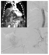
(A) CT angiography, coronal view: large right paravertebral thoracic mass with multiple arterial feeders (arrow). (B) The preliminary aortography demonstrates a rich vascularization of the tumor, mainly by branches from the VI, VIII, and X intercostal artery and an aberrant aortic branch (arrowhead). (CF) Selective angiography of the principal arterial feeders; note a radiculomedullary artery arising from the VIII intercostal artery (arrowhead in (C)).
Figure 2
Figure 2
(A) Superselective embolization with PVA particles and microcoils. (B) Final aortography shows devascularization of the mass.
Figure 3
Figure 3
(A) CT Angiography, coronal view: highly vascular mass inferior to the right hilum. Hypertrophic right phrenic artery ascending to the mass (arrow). (B) The preliminary aortography shows the presence of three hypertrophic bronchial feeders to the tumor with intralesional fistulae between bronchial arteries and the inferior pulmonary veins. (C,D) Selective angiogram of the superior right bronchial artery in different consecutive phases shows the recruitment of enlarged bronco-pulmonary anastomoses (arrowhead), with early opacification of the inferior pulmonary vein (arrow).
Figure 4
Figure 4
(A,B) The three bronchial feeders are superselectively microcatheterized and embolized with detachable coils. (C) Selective catheterization of the celiac trunk reveals hypertrophic right phrenic artery (arrow) feeding the tumor and communicating with the right inferior pulmonary vein (arrowhead).
Figure 5
Figure 5
(A) CT Angiography, coronal view: large, highly vascular mass in the left hemithorax dislodging the thoracic aorta. (B) Preliminary aortography shows the rich vascularization of the mass, with branches arising mainly from the left phrenic artery (arrow) and the first left lumbar artery (arrowhead). (C) Selective catheterization of the X intercostal left artery shows the presence of the Adamkiewicz artery (arrow) and a minimal supply to the tumor (arrowhead). (D) Highly reduced tumor vascularization at the final aortography.
Figure 6
Figure 6
(A) CT Angiography, coronal view: large mass in the posterior left mediastinum dislodging arterial pedicles arising from the thoracic aorta. (B) Preliminary aortography shows the mass has arterial feeders arising mainly from the left intercostal arteries. (C) Selective angiography of the VI intercostal left artery shows the presence of medial radicular branches (arrow). (D) Final aortography after PVA particle embolization.
Figure 7
Figure 7
(A) CT Angiography, coronal view: large mass in the posterior right hemithorax. Vascular supply from the right phrenic artery arising from the abdominal aorta (arrowhead). (B) Preliminary aortography shows hypertrophy of right intercostal arteries. (C) Selective angiography of the right phrenic artery and (D) right intercostobronchial trunk, with their supply to the tumor.
Figure 8
Figure 8
(A) Selective angiography of the VIII intercostal right artery demonstrates vascular supply to the tumor (arrow) and the anterior spinal artery of Adamkiewicz (arrowhead) arising from its radiculomedullary trunk. (B) Control after embolization demonstrates the preserved patency of the spinal artery (arrowhead). (C) Final aortography shows good devascularization of the tumor.

References

    1. Shi X., Liu X., Dong X., Wu H., Cai K. Trends, symptoms, and outcomes of resectable giant mediastinal tumors. Front. Oncol. 2022;12:820720. doi: 10.3389/fonc.2022.820720. - DOI - PMC - PubMed
    1. Sunam G.S., Öncel M., Ceran S., Ödev K., Yıldıran H. Giant benign mediastinal masses extending into the pleural cavity. Surg. J. 2016;2:e46–e50. doi: 10.1055/s-0036-1584519. - DOI - PMC - PubMed
    1. Darbari A., Dutt B., Kumar A., Goswami A.G., Starlet A.R. A retrospective observational study over surgical management of giant thoracic tumours: Horrendous but manageable. Cardiothorac. Surg. 2023;31:15. doi: 10.1186/s43057-023-00106-w. - DOI
    1. Madariaga M.L., Borges L.F., Rabinov J.D., Chang D.C., Lanuti M., Mathisen D.J., Gaissert H.A. Angiography before posterior mediastinal tumor resection: Selection criteria and patient outcomes. Ann. Thorac. Surg. 2018;105:1000–1007. doi: 10.1016/j.athoracsur.2017.12.028. - DOI - PubMed
    1. Brown M.A., Hu J., Tisol W., Grebe P., Howenstein M. Pre-operative embolization, surgical resection, and follow-up evaluation of a giant intercostal schwannoma. Clin. Imaging. 2022;85:74–77. doi: 10.1016/j.clinimag.2022.02.026. - DOI - PubMed

Publication types

LinkOut - more resources