. 2024 Oct 3;15(10):294.
doi: 10.3390/jfb15100294.

Nanofat Improves Vascularization and Tissue Integration of Dermal Substitutes without Affecting Their Biocompatibility

Affiliations

Nanofat Improves Vascularization and Tissue Integration of Dermal Substitutes without Affecting Their Biocompatibility

Francesca Bonomi et al. J Funct Biomater. .

Abstract

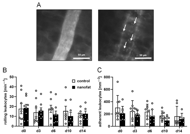
Dermal substitutes require sufficient tissue integration and vascularization to be successfully covered with split-thickness skin grafts. To rapidly achieve this, we provide the proof of principle for a novel vascularization strategy with high translational potential. Nanofat was generated from subcutaneous adipose tissue of green fluorescence protein (GFP)+ C57BL/6J donor mice and seeded onto small samples (4 mm in diameter) of the clinically approved dermal substitute Integra®. These samples and non-seeded controls were then implanted into full-thickness skin defects in the dorsal skinfold chamber of C57BL/6J wild-type mice and analyzed by intravital fluorescence microscopy, histology and immunohistochemistry over a 14-day period. Nanofat-seeded dermal substitutes exhibited an accelerated vascularization, as indicated by a significantly higher functional microvessel density on days 10 and 14 when compared to controls. This was primarily caused by the reassembly of GFP+ microvascular fragments inside the nanofat into microvascular networks. The improved vascularization promoted integration of the implants into the surrounding host tissue, which finally exhibited an increased formation of a collagen-rich granulation tissue. There were no marked differences in the inflammatory host tissue reaction to nanofat-seeded and control implants. These findings demonstrate that nanofat significantly improves the in vivo performance of dermal substitutes without affecting their biocompatibility.

Keywords: Integra®; angiogenesis; dermal substitutes; inflammation; nanofat; skin regeneration; vascularization.


Conflict of interest statement

The authors declare no conflicts of interest.

Figures

Figure 1
Figure 1
Methodological approach of this study. (A) Inguinal subcutaneous adipose tissue of a green fluorescence protein (GFP)+ donor mouse (border = broken line) with its inguinal lymph node (border = dotted line) for the generation of nanofat. (B) Three Luer-Lock connectors with descending internal diameters (2.4 mm, 1.4 mm, 1.2 mm) for mechanical fat emulsification. (C) Cell filter (pore size of 500 µm) sandwiched between two Luer-Lock connector halves for the filtration of the emulsified fat. (D) Freshly generated nanofat exhibiting a typical liquid consistency. (E) Sample that was cut out of a 1.3 mm thick, single-layer Integra® dermal regeneration template without a silicone sheet by means of a 4 mm biopsy punch. (F) Incubation of the sample in freshly generated nanofat. (G) Hematoxylin–eosin (HE)-stained cross-section of the sample after the seeding process. The nanofat is mainly located on the surface of the sample (asterisks). (H,I) Observation window of the dorsal skinfold chamber after implantation of a non-seeded (control, (H)) and nanofat-seeded (I) dermal substitute (border = broken line). (J) Schematic cross-section of a dorsal skinfold chamber with an implanted dermal substitute.
Figure 2
Figure 2
In vivo microscopy of dermal substitutes. (A) Stereomicroscopy of non-seeded (control, upper panels) and nanofat-seeded (lower panels) dermal substitutes on days 0, 3, 6, 10 and 14 (implant border = closed line). (B,C) Intravital fluorescence microscopy of non-seeded (control, (B)) and nanofat-seeded (C) dermal substitutes on days 0 and 14 (implant border = closed line; border of non-vascularized implant area = broken line). (DG) Perfused regions of interest (ROIs) (%) (D,E) and functional microvessel density (cm/cm2) (F,G) in the border (D,F) and center zones (E,G) of non-seeded (control; white bars, n = 8) and nanofat-seeded (black bars, n = 8) dermal substitutes on days 0, 3, 6, 10 and 14 after implantation, as assessed by intravital fluorescence microscopy. Means ± standard errors of the mean (SEMs). * p < 0.05 vs. control.
Figure 3
Figure 3
Leukocyte–endothelial cell interactions in response to dermal substitutes. (A) Intravital fluorescence microscopy of a collecting venule next to a dermal substitute (blue light epi-illumination, contrast enhancement by 5% fluorescein isothiocyanate (FITC)-labeled dextran (left panel); green light epi-illumination, in situ staining of leukocytes with 0.1% rhodamine 6G (right panel); arrows = leukocytes). (B,C) Rolling leukocytes (min−1) (B) and adherent leukocytes (mm−2) (C) within postcapillary and collecting venules in direct vicinity to non-seeded (control; white bars, n = 8) and nanofat-seeded (black bars, n = 8) dermal substitutes throughout the 14-day observation period. Means ± SEMs.
Figure 4
Figure 4
Tissue integration of dermal substitutes. (A,B) HE-stained sections of non-seeded (control, (A)) and nanofat-seeded (B) dermal substitutes on day 14 after implantation within dorsal skinfold chambers of C57BL/6J recipient mice (implant border = closed line; border zone = broken line; ROIs in the border and center zones of the implants shown in higher magnification in C and D = blue and red frame; panniculus carnosus muscle = arrows; granulation tissue = asterisks). (C,D) Higher magnification of blue and red frames in A and B in the border (C) and center (D) zones of the implants.
Figure 5
Figure 5
Vascularization and lymphatic drainage of dermal substitutes. (A) Immunohistochemical detection of CD31+ microvessels in the border zones (arrowheads) and the center (arrows) of non-seeded (control) and nanofat-seeded dermal substitutes on day 14 (implant border = closed line; border zones = dotted line). (B) Microvessel density (mm−2) of non-seeded (control; white bars, n = 8) and nanofat-seeded (black bars, n = 8) dermal substitutes on day 14, as assessed by immunohistochemistry. Means ± SEMs. * p < 0.05 vs. control. (C) Immunohistochemical detection of CD31+/GFP (arrows) and CD31+/GFP+ (arrowheads) microvessels in nanofat-seeded dermal substitutes on day 14. (D) CD31+/GFP+ microvessels (%) in the border zones and the center of nanofat-seeded dermal substitutes on day 14, as assessed by immunohistochemistry. Means ± SEMs. (E) Immunohistochemical detection of lymphatic vessel endothelial hyaluronan receptor (LYVE)-1+ lymph vessels in the border zones (arrowheads) and the center (arrow) of non-seeded (control) and nanofat-seeded dermal substitutes on day 14 (implant border = closed line; border zones = dotted line). (F) Lymph vessel density (mm−2) of non-seeded (control; white bars, n = 8) and nanofat-seeded (black bars, n = 8) dermal substitutes on day 14, as assessed by immunohistochemistry. Means ± SEMs. (G) Immunohistochemical detection of LYVE-1+/GFP- (arrow) and LYVE-1+/GFP+ (arrowheads) lymph vessels in nanofat-seeded dermal substitutes on day 14. (H) LYVE-1+/GFP+ microvessels (%) in the border zones and the center of nanofat-seeded dermal substitutes on day 14, as assessed by immunohistochemistry. Means ± SEMs.
Figure 6
Figure 6
Immune cell infiltration into dermal substitutes. (A,C,E) Immunohistochemical detection of CD68+ macrophages (A), myeloperoxidase (MPO)+ granulocytes (C) and CD3+ lymphocytes (E) in the border zones and the center of non-seeded (control) and nanofat-seeded dermal substitutes on day 14. (B,D,F) CD68+ macrophages (mm−2) (B), MPO+ granulocytes (mm−2) (D) and CD3+ lymphocytes (mm−2) (F) in the border zones and the center of non-seeded (control; white bars, n = 8) and nanofat-seeded (black bars, n = 8) dermal substitutes on day 14, as assessed by immunohistochemistry. Means ± SEMs. * p < 0.05 vs. control.
Figure 7
Figure 7
Collagen contents of dermal substitutes. (A,C) Immunohistochemical detection of collagen (Col) I (A) and III (C) in the border zones and the center of non-seeded (control) and nanofat-seeded dermal substitutes on day 14. (B,D) Total Col I (B) and Col III (D) ratio (implant/skin) in the border zones and the center of non-seeded (control; white bars, n = 8) and nanofat-seeded (black bars, n = 8) dermal substitutes on day 14, as assessed by immunohistochemistry. Means ± SEMs. * p < 0.05 vs. control.

References

    1. Eming S.A., Martin P., Tomic-Canic M. Wound repair and regeneration: Mechanisms, signaling, and translation. Sci. Transl. Med. 2014;6:265sr6. doi: 10.1126/scitranslmed.3009337. - DOI - PMC - PubMed
    1. Shevchenko R.V., James S.L., James S.E. A review of tissue-engineered skin bioconstructs available for skin reconstruction. J. R. Soc. Interface. 2010;7:229–258. doi: 10.1098/rsif.2009.0403. - DOI - PMC - PubMed
    1. Burke J.F., Yannas I.V., Quinby W.C., Bondoc C.C., Jung W.K. Successful use of a physiologically acceptable artificial skin in the treatment of extensive burn injury. Ann. Surg. 1981;194:413–428. doi: 10.1097/00000658-198110000-00005. - DOI - PMC - PubMed
    1. Reid M.J., Currie L.J., James S.E., Sharpe J.R. Effect of artificial dermal substitute, cultured keratinocytes and split thickness skin graft on wound contraction. Wound Repair Regen. 2007;15:889–896. doi: 10.1111/j.1524-475X.2007.00313.x. - DOI - PubMed
    1. Graham G.P., Helmer S.D., Haan J.M., Khandelwal A. The use of Integra® Dermal Regeneration Template in the reconstruction of traumatic degloving injuries. J. Burn. Care Res. 2013;34:261–266. doi: 10.1097/BCR.0b013e3182853eaf. - DOI - PubMed