. 2024 Oct 3;15(10):295.
doi: 10.3390/jfb15100295.

In Vivo Biocompatibility of Synechococcus sp. PCC 7002-Integrated Scaffolds for Skin Regeneration

Affiliations

In Vivo Biocompatibility of Synechococcus sp. PCC 7002-Integrated Scaffolds for Skin Regeneration

Benedikt Fuchs et al. J Funct Biomater. .

Abstract

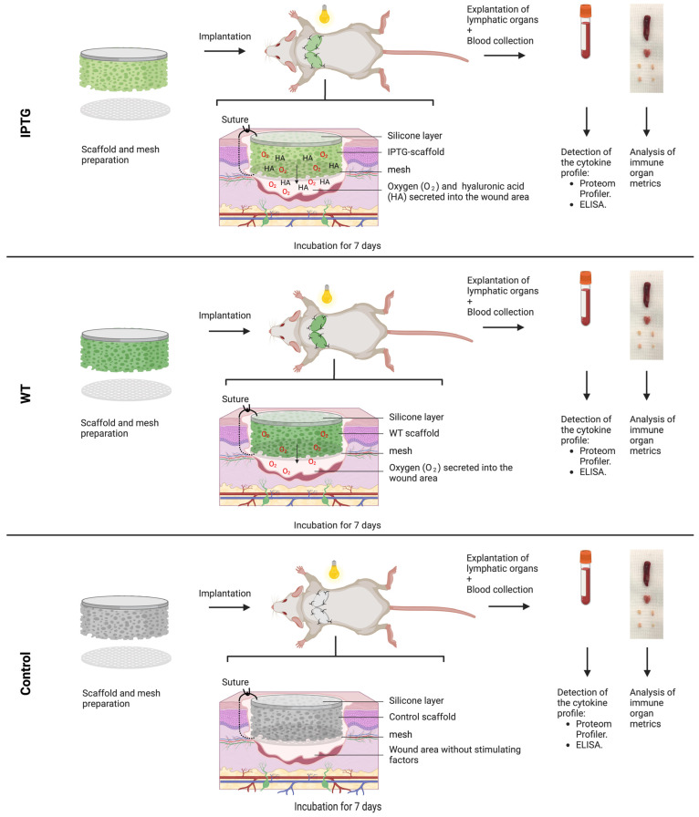
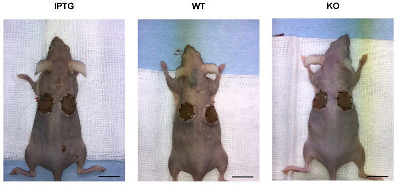
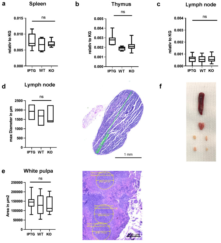
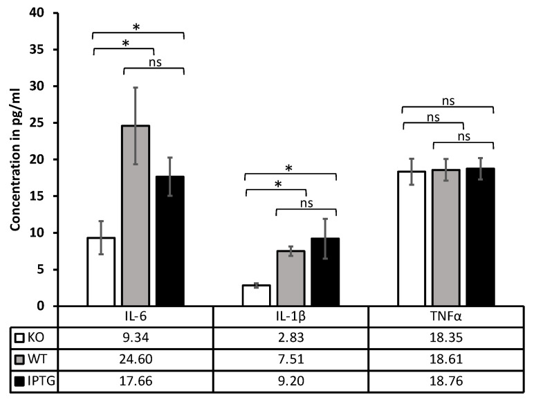
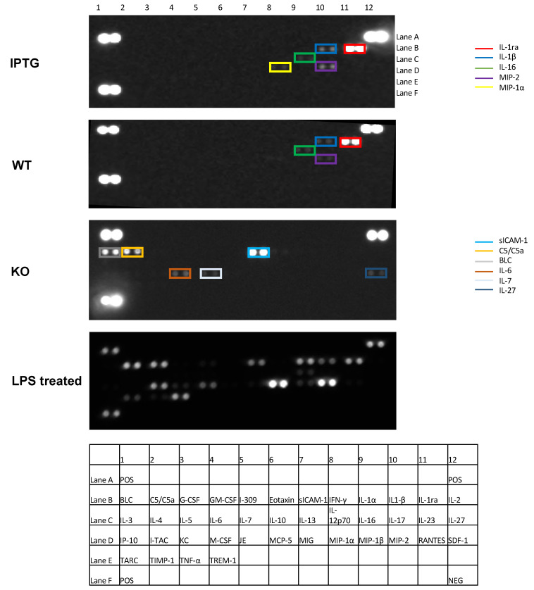
Cyanobacteria, commonly known as blue-green algae, are prevalent in freshwater systems and have gained interest for their potential in medical applications, particularly in skin regeneration. Among these, Synechococcus sp. strain PCC 7002 stands out because of its rapid proliferation and capacity to be genetically modified to produce growth factors. This study investigates the safety of Synechococcus sp. PCC 7002 when used in scaffolds for skin regeneration, focusing on systemic inflammatory responses in a murine model. We evaluated the following three groups: scaffolds colonized with genetically engineered bacteria producing hyaluronic acid, scaffolds with wild-type bacteria, and control scaffolds without bacteria. After seven days, we assessed systemic inflammation by measuring changes in cytokine profiles and lymphatic organ sizes. The results showed no significant differences in spleen, thymus, and lymph node weights, indicating a lack of overt systemic toxicity. Blood cytokine analysis revealed elevated levels of IL-6 and IL-1β in scaffolds with bacteria, suggesting a systemic inflammatory response, while TNF-α levels remained unaffected. Proteome profiling identified distinct cytokine patterns associated with bacterial colonization, including elevated inflammatory proteins and products, indicative of acute inflammation. Conversely, control scaffolds exhibited protein profiles suggestive of a rejection response, characterized by increased levels of cytokines involved in T and B cell activation. Our findings suggest that Synechococcus sp. PCC 7002 does not appear to cause significant systemic toxicity, supporting its potential use in biomedical applications. Further research is necessary to explore the long-term effects and clinical implications of these responses.

Keywords: Synechococcus sp. PCC 7002; biocompatibility; biomaterials; cyanobacteria; scaffolds; tissue engineering.


Conflict of interest statement

The authors declare no conflicts of interest.

Figures

Figure 1
Figure 1
Schematic illustration of the experimental protocol. Scaffolds with a diameter of 12 mm were inoculated with either IPTG-induced bacteria (IPTG) or wild-type bacteria (WT). A control group (KO) consisted of scaffolds devoid of bacteria. These prepared scaffolds were subsequently implanted alongside a mesh into bilateral full-thickness defects on the dorsal side of mice for a duration of seven days. Post-experimentation, the animals were euthanized, and blood samples were collected for the analysis of key inflammatory markers. Additionally, the lymphatic organs were excised and compared for variations in their dimensions.
Figure 2
Figure 2
Representative images of the mice of all 3 groups (IPGT, WT, and KO) using a stereoscope directly after euthanization by cervical vertebrae dislocation 7 days after scaffold implantation. The area of the implantation site is locally non-irritant without typical signs of inflammation such as redness, swelling, or hyperthermia. The scale bar represents 2 cm.
Figure 3
Figure 3
Analysis of immune organ metrics across different conditions. Demonstration of no systematic immune response 7 days after implantation of a scaffold colonized with IPTG-stimulated bacteria (IPTG), a scaffold with wild-type bacteria (WT), and a control scaffold (KO) in the absence of bacteria. We analyzed weight differences of the (a) spleen, (b) thymus, and (c) lymph nodes relative to the total weight of the mouse. (d) Left: We detected no significant differences in the maximal diameter of the axillar and inguinal lymph nodes. Right: Representative image of an inguinal lymph node in H&E staining. The scale bar represents 1 mm. (e) Left: The measurements of area in μm2 of the white pulp of the spleen revealed no marked differences in all groups. Right: Representative image of the white pulp in H&E staining. Magnification 5×. The scale bar represents 500 μm. Control scaffolds n = 10, colonized scaffolds n = 11, ns = not significant. (f) Representative image of the immune organs directly after explanation.
Figure 4
Figure 4
Detection of a systematic immune response 7 days after implantation of a scaffold colonized with IPTG-stimulated bacteria (IPTG), a scaffold with wild-type bacteria (WT), and a control scaffold (KO) in the absence of bacteria. Measurement of cytokine concentration in pg/mL by ELISA in mouse blood 7 days after scaffold implantation. The inflammatory cytokines detected are IL-6, IL-1β, and TNF-α. Cut off: IL-6 < 7.8 pg/mL, IL-1β < 6 pg/mL, TNF-α < 25 pg/mL, N ≥ 3; ns = not significant; * p < 0.5.
Figure 5
Figure 5
Detection of the cytokine profile 7 days after scaffold implantation in mouse blood using a proteome profiler. The following groups were analyzed: scaffolds with IPTG-stimulated bacteria (IPTG), scaffolds with wild-type bacteria (WT), control scaffolds (KO) in the absence of bacteria, and positive control (LPS treated) after LPS treatment. Significant differences exist in the cytokine profile between the scaffolds colonized with bacteria and the control scaffolds without bacteria. A table with the corresponding localization of the measured cytokines is attached. The positive control is shown in the top right, top left, and bottom left corners. The negative control is located at the bottom right. N > 3.

Similar articles

References

    1. Hamilton T.L., Bryant D.A., Macalady J.L. The role of biology in planetary evolution: Cyanobacterial primary production in low-oxygen Proterozoic oceans. Environ. Microbiol. 2016;18:325–340. doi: 10.1111/1462-2920.13118. - DOI - PMC - PubMed
    1. Rasala B.A., Mayfield S.P. Photosynthetic biomanufacturing in green algae; production of recombinant proteins for industrial, nutritional, and medical uses. Photosynth. Res. 2015;123:227–239. doi: 10.1007/s11120-014-9994-7. - DOI - PubMed
    1. Liu X., Xie H., Roussou S., Lindblad P. Current advances in engineering cyanobacteria and their applications for photosynthetic butanol production. Curr. Opin. Biotechnol. 2022;73:143–150. doi: 10.1016/j.copbio.2021.07.014. - DOI - PubMed
    1. Sarnaik A., Abernathy M.H., Han X., Ouyang Y., Xia K., Chen Y., Cress B., Zhang F., Lali A., Pandit R., et al. Metabolic engineering of cyanobacteria for photoautotrophic production of heparosan, a pharmaceutical precursor of heparin. Algal Res. 2019;37:57–63. doi: 10.1016/j.algal.2018.11.010. - DOI
    1. Li Z., Li S., Chen L., Sun T., Zhang W. Fast-growing cyanobacterial chassis for synthetic biology application. Crit. Rev. Biotechnol. 2024;44:414–428. doi: 10.1080/07388551.2023.2166455. - DOI - PubMed