Review
. 2024 Oct 18;10(10):724.
doi: 10.3390/jof10100724.

Overview of the Current Challenges in Pulmonary Coccidioidomycosis

Affiliations
Review

Overview of the Current Challenges in Pulmonary Coccidioidomycosis

Mohamed A Fayed et al. J Fungi (Basel). .

Abstract

Coccidioidomycosis is a disease caused by soil fungi of the genus Coccidioides, divided genetically into Coccidioides immitis (California isolates) and Coccidioides posadasii (isolates outside California). Coccidioidomycosis is transmitted through the inhalation of fungal spores, arthroconidia, which can cause disease in susceptible mammalian hosts, including humans. Coccidioidomycosis is endemic to the western part of the United States of America, including the central valley of California, Arizona, New Mexico, and parts of western Texas. Cases have been reported in other regions in different states, and endemic pockets are present in these states. The incidence of reported cases of coccidioidomycosis has notably increased since it became reportable in 1995. Clinically, the infection ranges from asymptomatic to fatal disease due to pneumonia or disseminated states. The recognition of coccidioidomycosis can be challenging, as it frequently mimics bacterial community-acquired pneumonia. The diagnosis of coccidioidomycosis is frequently dependent on serologic testing, the results of which can take several days or longer to obtain. Coccidioidomycosis continues to present challenges for clinicians, and suspected cases can be easily missed. The challenges of coccidioidomycosis disease, from presentation to diagnosis to treatment, remain a hurdle for clinicians, and further research is needed to address these challenges.

Keywords: coccidioidomycosis; lung mass; pulmonary nodules; pyopneumothorax; valley fever.


Conflict of interest statement

The author declares no conflicts of interest.

Figures
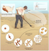
Figure 1
Figure 1
Saprobic life cycle of arthroconidia; the left-hand corner endemic map is from the CDC.
Figure 2
Figure 2
Life cycle of arthroconidia inside the lung, and typical parasitic forms of Coccidioides forming spherules and spherules/endospores.
Figure 3
Figure 3
T-cell immune response to fungal infection. Th1 and Th17 are critical for the elimination and effective control of dimorphic fungi infection such as Coccidioides.
Figure 4
Figure 4
Presentation of pulmonary of coccidioidomycosis.
Figure 5
Figure 5
(A) A chest X-ray shows a left lower opacity (blue arrow). (B) A CT scan shows left lower lobe consolidation (blue arrows). The patient was a 65-year-old woman who presented with the chief complaint of a cough and dyspnea lasting for one week. She was diagnosed with community-acquired pneumonia and placed on IV antibiotics and sent home with oral antibiotics. She tested positive for coccidioidomycosis IgG.
Figure 6
Figure 6
(A) shows a chest X-ray with left-sided pleural effusion (blue arrow). (B) shows a CT scan with left-sided pleural effusion (blue arrow). The patient was a 26-year-old woman who presented with progressive dyspnea and a cough associated with left-sided chest pain. She was originally diagnosed with community-acquired pneumonia until she presented to our hospital with worsening symptoms. She was diagnosed with pulmonary coccidioidomycosis via serology-positive IgM and her pleural fluid was positive for a coccidioidomycosis polymerase chain reaction (PCR).
Figure 7
Figure 7
(A) Pulmonary coccidioidomycosis presented with a lung mass (blue arrow). The patient was a 64-year-old man who presented with a cough, shortness of breath, and night sweats and his immunodiffusion serology was positive for coccidioidal IgM and IgG. (B) Pulmonary coccidioidomycosis presented with a lung mass (blue arrow). The patient was a 59-year-old man with a history of diabetes who presented with 3 days of a cough, malaise, night-sweats, and left upper back pain. He underwent a bronchoscopy and the bronchioalveolar lavage culture showed C. immitis.
Figure 8
Figure 8
(A) shows a large left upper lobe cavitary lesion. The patient was a 48-year-old woman who had cavitary disease discovered during a shoulder pain work-up (blue arrow). She later tested positive for IgM and IgG coccidioidomycosis serology. (B) shows a right lower lobe cavitary lesion. The patient was a 22-year-old man with a history of diabetes who presented with hemoptysis (blue arrow). The fungal culture from bronchial washing showed C. immitis.
Figure 9
Figure 9
(A) A chest X-ray shows diffuse reticulonodular interstitial miliary nodules (blue arrows). (B) A CT scan shows diffuse reticulonodular interstitial miliary nodules (blue arrows). The patient was a 27-year-old man with a history of human immunodeficiency virus (HIV) non-compliant on his HIV medication who presented with a duration of cough with yellow expectoration, night sweats, chills, severe weakness, and shortness of breath upon exertion. He was diagnosed with severe coccidioidomycosis via respiratory cultures and coccidioidomycosis fungemia.
Figure 10
Figure 10
(A) A chest X-ray shows pulmonary coccidioidomycosis mainly involving the right lung (blue arrow). (B) A CT scan shows pulmonary coccidioidomycosis mainly involving the right lung with consolidation (blue arrow). (C) A CT scan shows pulmonary coccidioidomycosis mainly involving the right lung with consolidation and evidence of necrosis (blue arrow). (D) A chest X-ray shows bilateral pulmonary infiltrates (red arrows) coccidioidomycosis with progression to severe ARDS (figure on day 7). The patient was placed on extracorporeal membrane oxygenation (ECMO) therapy (blue arrow shows ECMO cannula). The patient was a 37-year-old Filipino man with no medical history who presented with a fever, cough, and right lower chest pain for 2 weeks. He was initially admitted for CAP therapy, but his cardiorespiratory status deteriorated, necessitating extracorporeal membrane oxygenation (ECMO). Respiratory cultures grew Coccidioides spp.
Figure 11
Figure 11
(A) shows the initial chest X-ray with hydropneumothorax or pyopneumothorax, (effusion blue arrow and pneumothorax red arrow). (B) shows the initial CT scan with hydropneumothorax or pyopneumothorax (effusion blue arrow and consolidation red arrow). (C) shows the initial CT scan with hydropneumothorax or pyopneumothorax (effusion blue arrow and air red arrow). The patient was a 52-year-old male construction worker who presented with a progressive cough, shortness of breath, and left-sided chest pain. He was treated as having a traumatic hydropneumothorax, then diagnosed with complicated pulmonary coccidioidomycosis based on the histopathologic exam from his decortication specimen.
Figure 12
Figure 12
A CT scan with a left upper lobe nodule (blue arrow). The patient was a 64-year-old woman who presented with an incidental 2.5 cm left upper lobe nodule. She underwent a bronchoscopy with a biopsy, which was not diagnostic. Then, she underwent a CT guided biopsy, which showed necrotizing granuloma with fungal spherules consistent with coccidioidomycosis.
Figure 13
Figure 13
Areas of dissemination of coccidioidomycosis and/or associated symptoms.
Figure 14
Figure 14
(A) shows spherules of Coccidioides spp. in a pleural biopsy specimen stained with hematoxylin and eosin (blue arrow). (B) shows a Methenamine silver stain of a pleural biopsy specimen, showing spherules (blue arrow), endospores (black arrow), the rupture of spherules and release of endospores (red arrow), and hyphal forms (white arrow). The specimens were from a 52-year-old male construction worker who presented with a progressive cough, dyspnea, and left-sided chest pain. He was diagnosed with pyopneumothorax and underwent surgical thoracotomy and decortication. A histopathologic exam of the pleural biopsy specimen showed coccidioidomycosis spherules.
Figure 15
Figure 15
A left lower lung mass (blue arrow) that was suspected of malignancy. The mass has a subtle air bronchogram associated with ground-glass opacities and surrounding nodules. The patient was a 49-year-old patient who presented with night sweats, fatigue, and left-sided chest pain; her Coccidioides serologic testing for IgG was positive.
Figure 16
Figure 16
Proposed testing strategy for coccidioidomycosis. Cocci: coccidioidomycosis, EIA: enzyme immunoassay, LFA: lateral flow assay, ID: immunodiffusion, +ve: positive, −ve: negative.

References

    1. Benedict K., McCotter O.Z., Brady S., Komatsu K., Sondermeyer-Cooksey G.L., Nguyen A., Jain S., Vugia D.J., Jackson B.R. Surveillance for coccidioidomycosis—United States, 2011–2017. MMWR. 2019;68:1–15. doi: 10.15585/mmwr.ss6807a1. - DOI - PMC - PubMed
    1. Center for Disease Control and Prevention Valley Fever Map. [(accessed on 11 October 2024)]; Available online: https://www.cdc.gov/fungal/pdf/more-information-about-fungal-maps-508.pdf.
    1. Gorris M.E., Treseder K.K., Zender C.S., Randerson J.T. Expansion of Coccidioidomycosis Endemic Regions in the United States in Response to Climate Change. Geohealth. 2019;3:308–327. doi: 10.1029/2019GH000209. - DOI - PMC - PubMed
    1. Deresinski S., Mirels L.F. Coccidioidomycosis: What a long strange trip it’s been. Med. Mycol. 2019;57:S3–S15. doi: 10.1093/mmy/myy123. - DOI - PMC - PubMed
    1. Kirkland T.N., Stevens D.A., Hung C.-Y., Beyhan S., Taylor J.W., Shubitz L.F., Duttke S.H., Heidari A., Johnson R.H., Deresinski S.C., et al. Coccidioides Species: A Review of Basic Research: 2022. J. Fungi. 2022;8:859. doi: 10.3390/jof8080859. - DOI - PMC - PubMed