. 2024 Oct 2;13(10):866.
doi: 10.3390/pathogens13100866.

From Trypomastigotes to Trypomastigotes: Analyzing the One-Way Intracellular Journey of Trypanosoma cruzi by Ultrastructure Expansion Microscopy

Affiliations

From Trypomastigotes to Trypomastigotes: Analyzing the One-Way Intracellular Journey of Trypanosoma cruzi by Ultrastructure Expansion Microscopy

Ramiro Tomasina et al. Pathogens. .

Abstract

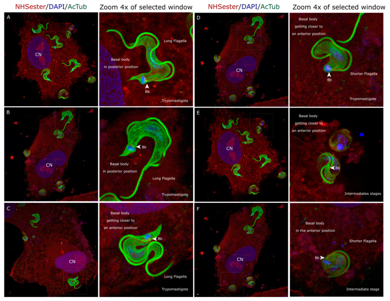
The protozoan parasite Trypanosoma cruzi is the causative agent of Chagas disease, also called American trypanosomiasis. This neglected tropical disease affects millions of individuals across the Americas. To complete its life cycle, T. cruzi parasitizes both vertebrate hosts and its vector, commonly known as the 'kissing bug'. The parasite's survival and proliferation strategies are driven by the diverse environments it encounters. Despite being described by Carlos Chagas in 1909, significant knowledge gaps persist regarding the parasite's various life forms and adaptive capabilities in response to environmental cues. In this study, we employed Ultrastructure Expansion Microscopy to explore the intricate journey of T. cruzi within the host cell. Upon entry into the host cell, trypomastigotes undergo folding, resulting in intermediate forms characterized by a rounded cell body, anterior positioning of basal bodies, and a shortened flagellum. The repositioning of basal bodies and the kinetoplast and the shortening of the flagella mark the culmination of intracellular amastigogenesis. Furthermore, we analyzed intracellular trypomastigogenesis, identifying discrete intermediate forms, including leaf-shaped stages and epimastigote-like forms, which suggests a complex differentiation process. Notably, we did not observe any dividing intracellular epimastigotes, indicating that these may be non-replicative forms within the host cell. Our detailed examination of amastigote cell division revealed semi-closed nuclear mitosis, with mitotic spindle formation independent of basal bodies. This study provides new insights into the morphological and cytoskeletal changes during the intracellular stages of T. cruzi, providing a model for understanding the dynamics of intracellular amastigogenesis and trypomastigogenesis.

Keywords: Trypanosoma cruzi; UExM; amastigogenesis; cell division; trypomastigogenesis.


Conflict of interest statement

The authors declare no conflict of interest.

Figures

Figure 1
Figure 1
Intracellular trypomastigotes and rounded forms 30 min post-infection. (AC) Trypomastigotes observed under UExM inside the cell. (C) Intermediate rounded shape form observed by UExM inside the cell. (D,E) In this rolling process, the parasite’s posterior part comes closer to its anterior part, making possible the position of the basal body and kinetoplast observed in the amastigotes. (F) In this panel, an intermediate rounded-shape life form is observed. Although similar to an amastigote form, it holds a longer flagellum. ‘Bb’ stands for basal bodies; ‘CN’ stands for cell nucleus. All the images are maximum-intensity projection z-stacks. The scale bar is 1 µm in all images.
Figure 2
Figure 2
Intracellular amastigogenesis. (A) UExM of selected different life forms observed at 30 min post-infection and 4 and 24 h post-infection. All these life forms were observed in the process of the observation of amastigotes. (B) Amastigogenesis model. Upon entering the cell, the trypomastigotes undergo several transformations. These include shortening of the flagella and repositioning of the basal body and kinetoplast towards the nucleus. We propose a mechanism where the parasite executes a rolling movement, reduces its cell body size, and brings its posterior part in contact with the anterior part. Through a process of membrane fusion, T. cruzi forms a single enlarged rounded cell body, which subsequently gives rise to the amastigotes.
Figure 3
Figure 3
Three dividing forms are observed at 24 h post-infection. Using UExM, we could observe in detail the intracellular life form present at 24 h post-infection. Interestingly, although we assumed all of them were amastigotes, significant structural differences were observed. In the first row, we can observe that some of these lifeforms hold very short flagella and are perfectly rounded shapes. In the second row, we can observe rounded shape lifeforms but with longer flagella. In the third row, we show another life form observed that holds a leaf shape with short flagella. The columns of intracellular forms feature selected images in which the background and surroundings of the parasite have been removed, enhancing the visibility of the parasite’s morphology. All these life forms are replicative and with short flagella, which aligns with the definition of amastigotes. In rows 2 and 3, the helical arrangement of microtubules from the cytostome cytoskeleton is observed (yellow arrows). ‘Bb’ stands for basal bodies; ‘CN’ stands for cell nucleus. All the images are maximum-intensity projections of selected z-stacks to allow for the visualization of the structures pointed. The scale bar is 1 µm.
Figure 4
Figure 4
(AL) Intracellular forms 72 h post-infection. At 72 h post-infection, not only amastigotes and trypomastigotes are observed. Consistent with previous observations, we observe epimastigote-like life forms, a ‘drop epimastigote’-like form and many intermediate amastigote forms, which meet the definition of amastigotes because of the position of the basal body, the kinetoplast, and nucleus. Still, they have a more sharpened shape instead of the rounded shape. All the images are maximum-intensity projections of selected z-stacks to allow for the visualization of the structures pointed. Please note that the columns of intracellular forms are selected images that have been cut using Inkscape V 1.3.2 (imaging software) to clarify their morphology observation. The helical arrangement of microtubules from the cytostome–cytopharynx cytoskeleton is indicated by yellow arrows. ‘Bb’ stands for basal bodies; ‘CN’ stands for cell nucleus. Please note that at 72 h, the number of parasites inside the cells was huge, and the DAPI signal tended to be faint and messy, making the visualization of the parasites difficult, which is why we decided not to include them in the figure. The scale bar is 1 µm in all images.
Figure 5
Figure 5
Amastigote cell division needs the assembly of a mitotic spindle nucleated without centrioles. During division, amastigotes assemble the mitotic spindle without disassembling the nuclear membrane and nucleating them without centrioles. Using UExM, we could observe in detail the binary fission process as never shown by immunofluorescence microscopy. Images were taken at 24 h post-infection. All the images are maximum-intensity projections of selected z-stacks to allow for the visualization of the structures indicated. The scale bar is 1 µm in all images.
Figure 6
Figure 6
Intracellular trypomastigogenesis. (A) UExM of selecting different life forms observed 72 h post-infection. (B) Trypomastigogenesis model. As previously described, T. cruzi suffers several changes from amastigote to trypomastigote inside the infected cell. Using UExM, we could observe for the first time all these intermediate forms in detail that had never been obtained before using an immunofluorescence technique. The resolution achieved enabled us to identify two previously undescribed intermediate forms: one between the transition from amastigote to epimastigote and another between the transformation of epimastigotes into trypomastigotes, as illustrated in panels (A) and (B).
Figure 7
Figure 7
Intracellular life stages of Trypanosoma cruzi by UExM. UExM of selecting different life forms observed at 30 min, 4, 24, and 72 h post-infection. All the intracellular forms are selected images that have been cut using Inkscape V 1.3.2 (imaging software) to clarify the morphology observation.

References

    1. Chagas C. Nova tripanozomiaze humana: Estudos sobre a morfolojia e o ciclo evolutivo do Schizotrypanum cruzi n. gen., n. sp., ajente etiolojico de nova entidade morbida do homem. Mem. Inst. Oswaldo Cruz. 1909;1:159–218. doi: 10.1590/S0074-02761909000200008. - DOI
    1. Vickerman K. The Diversity of the Kinetoplastid Flagellates. 1977. [(accessed on 29 September 2024)]. Available online: https://www.cabidigitallibrary.org/doi/full/10.5555/19762501581. - DOI
    1. De Souza W. Cell biology of Trypanosoma cruzi. Int. Rev. Cytol. 1984;86:197–283. doi: 10.1016/s0074-7696(08)60180-1. - DOI - PubMed
    1. Gonçalves C.S., Ávila A.R., De Souza W., Motta M.C.M., Cavalcanti D.P. Revisiting the Trypanosoma cruzi metacyclogenesis: Morphological and ultrastructural analyses during cell differentiation. Parasites Vectors. 2018;11:83. doi: 10.1186/s13071-018-2664-4. - DOI - PMC - PubMed
    1. Vidal J.C., De Souza W. 3D FIB-SEM structural insights into the architecture of sub-pellicular microtubules of Trypanosoma cruzi epimastigotes. Biol. Cell. 2022;114:203–210. doi: 10.1111/boc.202100038. - DOI - PubMed

LinkOut - more resources