. 2024 Oct 4;22(10):456.
doi: 10.3390/md22100456.

Potential of Marine Sponge Metabolites against Prions: Bromotyrosine Derivatives, a Family of Interest

Affiliations

Potential of Marine Sponge Metabolites against Prions: Bromotyrosine Derivatives, a Family of Interest

Maha Sinane et al. Mar Drugs. .

Abstract

The screening of 166 extracts from tropical marine organisms (invertebrates, macroalgae) and 3 cyclolipopeptides from microorganisms against yeast prions highlighted the potential of Verongiida sponges to prevent the propagation of prions. We isolated the known compounds purealidin Q (1), aplysamine-2 (2), pseudoceratinine A (3), aerophobin-2 (4), aplysamine-1 (5), and pseudoceratinine B (6) for the first time from the Wallisian sponge Suberea laboutei. We then tested compounds 1-6 and sixteen other bromotyrosine and bromophenol derivatives previously isolated from Verongiida sponges against yeast prions, demonstrating the potential of 1-3, 5, 6, aplyzanzine C (7), purealidin A (10), psammaplysenes D (11) and F (12), anomoian F (14), and N,N-dimethyldibromotyramine (15). Following biological tests on mammalian cells, we report here the identification of the hitherto unknown ability of the six bromotyrosine derivatives 1, 2, 5, 7, 11, and 14 of marine origin to reduce the spread of the PrPSc prion and the ability of compounds 1 and 2 to reduce endoplasmic reticulum stress. These two biological activities of these bromotyrosine derivatives are, to our knowledge, described here for the first time, offering a new therapeutic perspective for patients suffering from prion diseases that are presently untreatable and consequently fatal.

Keywords: ER stress; PrPSc; Suberea laboutei; Verongiida; bromotyrosine derivatives; marine sponges; prion diseases; yeast-based screening.


Conflict of interest statement

The authors declare no conflicts of interest. The funders had no role in the design of the study; in the collection, analyses, or interpretation of data; in the writing of the manuscript; or in the decision to publish the results.

Figures

Figure 1
Figure 1
Sponge extracts displaying activity against [PSI+] and [URE3] yeast prions. Yeast cells were spread on a rich agar medium and small sterile filters were then placed on the agar surface, and 2 µL of each 10 mg/mL sponge extract was applied to the individual filters. A red halo around the filter on which an extract was loaded indicates that the extract is active against [PSI+] or [URE3] prions, whereas colonies that remain white indicate inactive extracts. Screening scores were attributed according to Table 1.
Figure 2
Figure 2
Structures of the isolated compounds 16 from Suberea laboutei.
Figure 3
Figure 3
Anti-prion activity of compounds 16 against [PSI+] and [URE3] yeast prions as described in Figure 1. Screening scores were attributed according to Table 1. Molecules 1, 2, 3, 5, and 6 are active against both yeast prions.
Figure 4
Figure 4
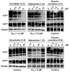
Anti-PrPSc activity of compounds 16 on prion-infected MovS6 cells. MovS6 cells were treated with the indicated ranges of concentrations of the molecules, and absolute ethanol was used as a negative control. After 6 days of culture, cell lysates were digested by proteinase K to reveal PrPSc (top panels) or untreated to reveal PrPC (middle panels) or the loading control Tubulin (bottom panels). Proteins were separated in 10% Bis-Tris polyacrylamide gels and revealed using anti-PrP (Sha31) or anti-tubulin-specific antibodies. The blots shown are representative of two to three independent experiments that all produced similar results. Purealidin Q (1), aplysamine-2 (2) and aplysamine-1 (5) were able to reduce PrPSc propagation, whereas pseudoceratinine A (3), aerophobin-2 (4), and pseudoceratinine-B (6) were not.
Figure 5
Figure 5
Structures of the tested bromotyrosine and bromophenol derivatives 722.
Figure 6
Figure 6
Activity of bromotyrosine derivatives 722 against [PSI+] (a) and [URE3] (b) yeast prions, as described in Figure 1. Screening scores were attributed according to Table 1. Compounds 7, 10, 11, 12, 14, and 15 were active against both [PSI+] and [URE3] yeast prions.
Figure 7
Figure 7
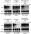
Activity of bromotyrosine derivatives 7, 10, 11, 12, 14, and 15 against the PrPSc prion and on prion-infected MovS6 cells, as described in Figure 4. The blots shown are representative of two to three independent experiments that all produced similar results. Aplyzanzine C (7), psammaplysene D (11), and anomoian F (14) were able to reduce PrPSc propagation, whereas purealidin A (10), psammaplysene F (12) and N,N-dimethyl-dibromotyramine (15) were not.
Figure 8
Figure 8
Cytoprotective activity of bromotyrosine derivatives purealidin Q (1), aplysamine-2 (2), aplysamine-1 (5), aplyzanzine C (7), psammaplysene D (11), and anomoian F (14) against ER stress. CHO-K1 cells were ER-stressed using 0.45 µg/mL tunicamycin (Tm) in the presence of the indicated concentrations of the test molecules or DMSO as a negative control. After 24 h of treatment, cell viability was measured by the quantification of the number of live cells using the WST-8 tetrazolium salt. Values are shown relative to the DMSO-treated cells, which was set at a value of 100%. A representative assay including three technical repeats is shown with SD error bars, and each experiment was performed at least three times with similar results. Bar height represents the mean relative to DMSO-treated cells. **** p < 0.0001 one-way ANOVA compared with Tm-treated cells, followed by Dunnett’s test.
Figure 9
Figure 9
The effect of purealidin Q (1) and aplysamine-2 (2) on CHOP expression level. After 24 h treatment of CHO-K1 cells with Tm and test molecules as described in Figure 8, luciferase activity was measured. A representative assay including three technical repeats is shown with SD error bars, and each experiment was performed at least three times with similar results. Bar height represents the mean relative to DMSO-treated cells. ** p < 0.01, **** p < 0.0001 one-way ANOVA compared with Tm-treated cells, followed by Dunnett’s test.

References

    1. Griffith J.S. Nature of the Scrapie Agent: Self-Replication and Scrapie. Nature. 1967;215:1043–1044. doi: 10.1038/2151043a0. - DOI - PubMed
    1. Prusiner S.B. Novel Proteinaceous Infectious Particles Cause Scrapie. Science. 1982;216:136–144. doi: 10.1126/science.6801762. - DOI - PubMed
    1. Prusiner S.B. Prions. Proc. Natl. Acad. Sci. USA. 1998;95:13363–13383. doi: 10.1073/pnas.95.23.13363. - DOI - PMC - PubMed
    1. Collinge J. Mammalian Prions and Their Wider Relevance in Neurodegenerative Diseases. Nature. 2016;539:217–226. doi: 10.1038/nature20415. - DOI - PubMed
    1. Scheckel C., Aguzzi A. Prions, Prionoids and Protein Misfolding Disorders. Nat. Rev. Genet. 2018;19:405–418. doi: 10.1038/s41576-018-0011-4. - DOI - PubMed

LinkOut - more resources