. 2024 Nov;6(6):e240073.
doi: 10.1148/rycan.240073.

Deformable Mapping of Rectal Cancer Whole-Mount Histology with Restaging MRI at Voxel Scale: A Feasibility Study

Affiliations

Deformable Mapping of Rectal Cancer Whole-Mount Histology with Restaging MRI at Voxel Scale: A Feasibility Study

João Miranda et al. Radiol Imaging Cancer. 2024 Nov.

Abstract

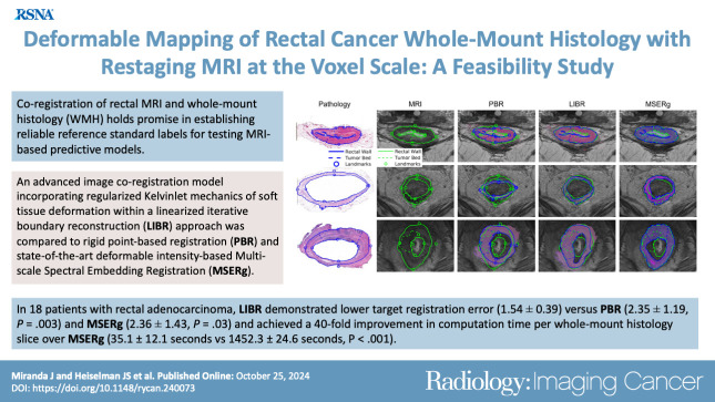
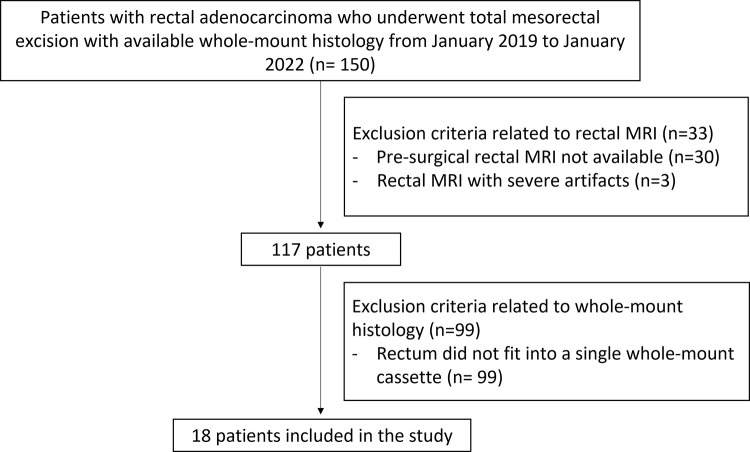
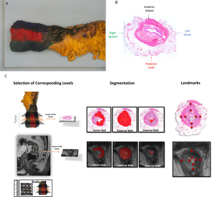
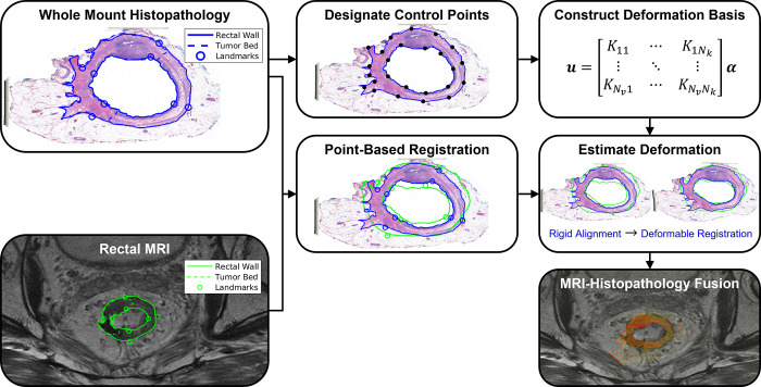
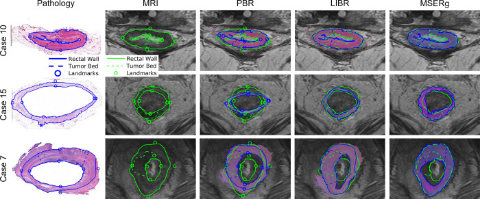
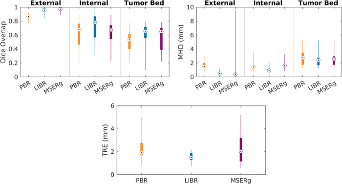
Purpose To develop a radiology-pathology coregistration method for 1:1 automated spatial mapping between preoperative rectal MRI and ex vivo rectal whole-mount histology (WMH). Materials and Methods This retrospective study included consecutive patients with rectal adenocarcinoma who underwent total neoadjuvant therapy followed by total mesorectal excision with preoperative rectal MRI and WMH from January 2019 to January 2022. A gastrointestinal pathologist and a radiologist established three corresponding levels for each patient at rectal MRI and WMH, subsequently delineating external and internal rectal wall contours and the tumor bed at each level and defining eight point-based landmarks. An advanced deformable image coregistration model based on the linearized iterative boundary reconstruction (LIBR) approach was compared with rigid point-based registration (PBR) and state-of-the-art deformable intensity-based multiscale spectral embedding registration (MSERg). Dice similarity coefficient (DSC), modified Hausdorff distance (MHD), and target registration error (TRE) across patients were calculated to assess the coregistration accuracy of each method. Results Eighteen patients (mean age, 54 years ± 13 [SD]; nine female) were included. LIBR demonstrated higher DSC versus PBR for external and internal rectal wall contours and tumor bed (external: 0.95 ± 0.03 vs 0.86 ± 0.04, respectively, P < .001; internal: 0.71 ± 0.21 vs 0.61 ± 0.21, P < .001; tumor bed: 0.61 ± 0.17 vs 0.52 ± 0.17, P = .001) and versus MSERg for internal rectal wall contours (0.71 ± 0.21 vs 0.63 ± 0.18, respectively; P < .001). LIBR demonstrated lower MHD versus PBR for external and internal rectal wall contours and tumor bed (external: 0.56 ± 0.25 vs 1.68 ± 0.56, respectively, P < .001; internal: 1.00 ± 0.35 vs 1.62 ± 0.59, P < .001; tumor bed: 2.45 ± 0.99 vs 2.69 ± 1.05, P = .03) and versus MSERg for internal rectal wall contours (1.00 ± 0.35 vs 1.62 ± 0.59, respectively; P < .001). LIBR demonstrated lower TRE (1.54 ± 0.39) versus PBR (2.35 ± 1.19, P = .003) and MSERg (2.36 ± 1.43, P = .03). Computation time per WMH slice for LIBR was 35.1 seconds ± 12.1. Conclusion This study demonstrates feasibility of accurate MRI-WMH coregistration using the advanced LIBR method. Keywords: MR Imaging, Abdomen/GI, Rectum, Oncology Supplemental material is available for this article. © RSNA, 2024.

Keywords: Abdomen/GI; MR Imaging; Oncology; Rectum.


Conflict of interest statement

Disclosures of conflicts of interest: J.M. No relevant relationships. J.S.H. No relevant relationships. C.F. No relevant relationships. J.C. No relevant relationships. R.S.V. No relevant relationships. A.N.A. No relevant relationships. J.N. Financial support from Memorial Sloan Kettering Cancer Center to present at and attend the 2024 European Congress of Radiology. T.H.K. No relevant relationships. L.R. No relevant relationships. N.U. No relevant relationships. M.G. Support from the National Institutes of Health (grant no. P30 CA008748), paid to author’s employer. J.G.A. Stock in Intuitive Surgical. M.J.G. No relevant relationships. J.S. No relevant relationships. N.H. One-time consultant agreement with Guerbet (February 2023); one-time speaker agreement with Bayer (September 2022) and Guerbet (May 2024); travel support from Guerbet (February 2023 for Society of Abdominal Radiology and May 2024 for Jornada Paulista de Radiologia).

Figures

None
Graphical abstract
Flowchart of patient inclusion. This retrospective study included
consecutive patients with rectal adenocarcinoma who underwent total
mesorectal excision at our institution from January 2019 to January 2022 and
who had available whole-mount histology (WMH) specimens. The exclusion
criteria were as follows: WMH slices not fitting into a single whole-mount
cassette, presurgical MRI data unavailable, and severe restaging MRI
artifacts.
Figure 1:
Flowchart of patient inclusion. This retrospective study included consecutive patients with rectal adenocarcinoma who underwent total mesorectal excision at our institution from January 2019 to January 2022 and who had available whole-mount histology (WMH) specimens. The exclusion criteria were as follows: WMH slices not fitting into a single whole-mount cassette, presurgical MRI data unavailable, and severe restaging MRI artifacts.
Demarcation of a standard total mesorectal excision specimen on (A, B)
surgical and whole-mount histology (WMH) slices and (C) radiology-pathology
workflow of segmentation and landmark definition. (A) Circumferential
margins of the rectal cancer resection specimen were marked with different
ink colors to distinguish anterior and posterior and right and left regions
of the specimen and enable proper orientation. (B) WMH slice of the total
mesorectal excision specimen demonstrates the color code that was used to
guide the spatial localization of the rectum portions, as follows: black =
anterior, red = posterior, blue = left, and green = right. (C) A
gastrointestinal pathologist and a radiologist conducted a collaborative
review of WMH and MR images to ensure precise correspondence between
pathology and high-resolution T2-weighted imaging. Three corresponding
levels were established for each patient at WMH and MRI: the midpoint of the
tumor bed, one slice or section above, and one slice or section below.
Subsequently, both experts manually delineated the external rectal contour
(the outer edge of the muscularis propria), internal rectal contour (inner
aspect of the mucosa), and tumor bed at each designated level (illustrated
at the midpoint level). Additionally, the radiologist and pathologist
annotated eight corresponding point-based landmarks in each modality along
the internal and external borders of the rectal wall, encompassing the
anterior, posterior, leftward, and rightward directions.
Figure 2:
Demarcation of a standard total mesorectal excision specimen on (A, B) surgical and whole-mount histology (WMH) slices and (C) radiology-pathology workflow of segmentation and landmark definition. (A) Circumferential margins of the rectal cancer resection specimen were marked with different ink colors to distinguish anterior and posterior and right and left regions of the specimen and enable proper orientation. (B) WMH slice of the total mesorectal excision specimen demonstrates the color code that was used to guide the spatial localization of the rectum portions, as follows: black = anterior, red = posterior, blue = left, and green = right. (C) A gastrointestinal pathologist and a radiologist conducted a collaborative review of WMH and MR images to ensure precise correspondence between pathology and high-resolution T2-weighted imaging. Three corresponding levels were established for each patient at WMH and MRI: the midpoint of the tumor bed, one slice or section above, and one slice or section below. Subsequently, both experts manually delineated the external rectal contour (the outer edge of the muscularis propria), internal rectal contour (inner aspect of the mucosa), and tumor bed at each designated level (illustrated at the midpoint level). Additionally, the radiologist and pathologist annotated eight corresponding point-based landmarks in each modality along the internal and external borders of the rectal wall, encompassing the anterior, posterior, leftward, and rightward directions.
Coregistration workflow for the proposed linearized iterative boundary
reconstruction (LIBR) MRI-histopathology fusion method after segmentation
and landmark characterization as demonstrated in Figure 2. Prior to
coregistration, eight point-based landmarks are annotated on the external
and internal rectal contours on each MR and whole-mount histopathology (WMH)
image. A series of regularized Kelvinlet control points is then distributed
across the external and internal rectal contours, from which a biomechanical
deformation basis is constructed. After point-based registration of
landmarks, deformation of the histopathology sample relative to MRI is
computed from the rectal wall contours via the LIBR approach. A series of
control points are distributed across the external and internal rectal
contours to establish a regularized Kelvinlet deformation basis for the WMH
image. The LIBR approach estimates the deformation between WMH and MRI by
maximizing the agreement between rectal wall contours subject to this
deformation basis. Finally, the registered WMH is fused with MRI to indicate
spatial correspondence between reference standard histopathologic features
and MRI.
Figure 3:
Coregistration workflow for the proposed linearized iterative boundary reconstruction (LIBR) MRI-histopathology fusion method after segmentation and landmark characterization as demonstrated in Figure 2. Prior to coregistration, eight point-based landmarks are annotated on the external and internal rectal contours on each MR and whole-mount histopathology (WMH) image. A series of regularized Kelvinlet control points is then distributed across the external and internal rectal contours, from which a biomechanical deformation basis is constructed. After point-based registration of landmarks, deformation of the histopathology sample relative to MRI is computed from the rectal wall contours via the LIBR approach. A series of control points are distributed across the external and internal rectal contours to establish a regularized Kelvinlet deformation basis for the WMH image. The LIBR approach estimates the deformation between WMH and MRI by maximizing the agreement between rectal wall contours subject to this deformation basis. Finally, the registered WMH is fused with MRI to indicate spatial correspondence between reference standard histopathologic features and MRI.
Visualization of the alignment quality achieved by each coregistration
method. Restaging MR–whole-mount histology image fusion is shown in
three representative cases. Case 10 (a 43-year-old female patient), case 15
(a 65-year-old male patient), and case 7 (a 47-year-old male patient)
represent cases with the best Dice similarity coefficient, average Dice
similarity coefficient, and worst Dice similarity coefficient, respectively.
Between the investigated linearized iterative boundary reconstruction
(LIBR), rigid point-based registration (PBR), and multiscale spectral
embedding registration (MSERg) methods, LIBR produced better alignment. Note
that in case 7, MSERg produced a misalignment of approximately 90°
between the pathology slice and the MR image.
Figure 4:
Visualization of the alignment quality achieved by each coregistration method. Restaging MR–whole-mount histology image fusion is shown in three representative cases. Case 10 (a 43-year-old female patient), case 15 (a 65-year-old male patient), and case 7 (a 47-year-old male patient) represent cases with the best Dice similarity coefficient, average Dice similarity coefficient, and worst Dice similarity coefficient, respectively. Between the investigated linearized iterative boundary reconstruction (LIBR), rigid point-based registration (PBR), and multiscale spectral embedding registration (MSERg) methods, LIBR produced better alignment. Note that in case 7, MSERg produced a misalignment of approximately 90° between the pathology slice and the MR image.
Distribution of accuracy values for each coregistration method across
the study sample. Coregistration accuracy was assessed for our linearized
iterative boundary reconstruction (LIBR) method, the rigid point-based
registration (PBR) method, and the multiscale spectral embedding
registration (MSERg) method using the Dice similarity coefficient (DSC), the
modified Hausdorff distance (MHD), and the target registration error (TRE).
The LIBR method produced lower TRE, DSC, and MHD than the rigid PBR method
across all features evaluated (external rectal contour, internal rectal
contour, and tumor bed) and also produced lower TRE, DSC, and MHD than MSERg
along the internal rectal wall.
Figure 5:
Distribution of accuracy values for each coregistration method across the study sample. Coregistration accuracy was assessed for our linearized iterative boundary reconstruction (LIBR) method, the rigid point-based registration (PBR) method, and the multiscale spectral embedding registration (MSERg) method using the Dice similarity coefficient (DSC), the modified Hausdorff distance (MHD), and the target registration error (TRE). The LIBR method produced lower TRE, DSC, and MHD than the rigid PBR method across all features evaluated (external rectal contour, internal rectal contour, and tumor bed) and also produced lower TRE, DSC, and MHD than MSERg along the internal rectal wall.

References

    1. Sung H , Ferlay J , Siegel RL , et al . Global Cancer Statistics 2020: GLOBOCAN Estimates of Incidence and Mortality Worldwide for 36 Cancers in 185 Countries . CA Cancer J Clin 2021. ; 71 ( 3 ): 209 – 249 . - PubMed
    1. Siegel RL , Torre LA , Soerjomataram I , et al . Global patterns and trends in colorectal cancer incidence in young adults . Gut 2019. ; 68 ( 12 ): 2179 – 2185 . - PubMed
    1. Vuik FE , Nieuwenburg SA , Bardou M , et al . Increasing incidence of colorectal cancer in young adults in Europe over the last 25 years . Gut 2019. ; 68 ( 10 ): 1820 – 1826 . - PMC - PubMed
    1. Kong JC , Soucisse M , Michael M , et al . Total neoadjuvant therapy in locally advanced rectal cancer: a systematic review and metaanalysis of oncological and operative outcomes . Ann Surg Oncol 2021. ; 28 ( 12 ): 7476 – 7486 . - PubMed
    1. Garcia-Aguilar J , Patil S , Gollub MJ , et al . Organ preservation in patients with rectal adenocarcinoma treated with total neoadjuvant therapy . J Clin Oncol 2022. ; 40 ( 23 ): 2546 – 2556 . - PMC - PubMed