Review
. 2024 Oct 14;14(20):1648.
doi: 10.3390/nano14201648.

Introduction and Development of Surface-Enhanced Raman Scattering (SERS) Substrates: A Review

Affiliations
Review

Introduction and Development of Surface-Enhanced Raman Scattering (SERS) Substrates: A Review

Jianping Peng et al. Nanomaterials (Basel). .

Abstract

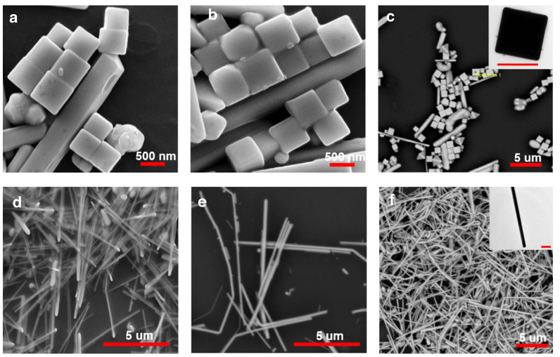
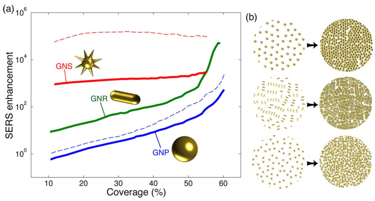
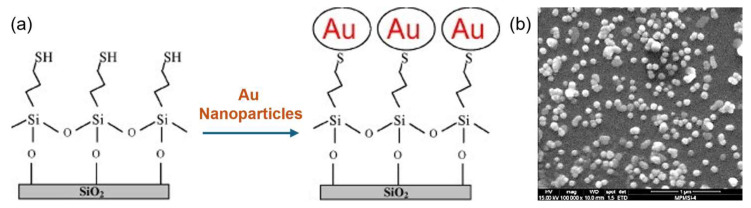
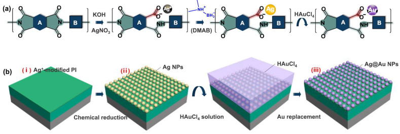
Since its discovery, the phenomenon of Surface Enhanced Raman Scattering (SERS) has gradually become an important tool for analyzing the composition and structure of substances. As a trace technique that can efficiently and nondestructively detect single molecules, the application of SERS has expanded from environmental and materials science to biomedical fields. In the past decade or so, the explosive development of nanotechnology and nanomaterials has further boosted the research of SERS technology, as nanomaterial-based SERS substrates have shown good signal enhancement properties. So far, it is widely recognized that the morphology, size, composition, and stacking mode of nanomaterials have a very great influence on the strength of the substrate SERS effect. Herein, an overview of methods for the preparation of surface-enhanced Raman scattering (SERS) substrates is provided. Specifically, this review describes a variety of common SERS substrate preparation methods and explores the potential and promise of these methods for applications in chemical analysis and biomedical fields. By detailing the influence of different nanomaterials (e.g., metallic nanoparticles, nanowires, and nanostars) and their structural features on the SERS effect, this article aims to provide a comprehensive understanding of SERS substrate preparation techniques.

Keywords: SERS; preparation; substrates.


Conflict of interest statement

The authors declare no conflict of interest.

Figures

Figure 1
Figure 1
SEM images (ac) silver nanocubes (df) silver nanowires SEM images (ac) silver nanocubes (df) silver nanowires (the inset shows the TEM micrograph of corresponding nanocube and nanowire with a scale bar of 500 nm) [57]. Copyright © 2020, Springer.
Figure 2
Figure 2
Density dependence of the SERS performance in nanoparticle monolayers. The particle coverage is defined as the fraction of area occupied by the projection of the metal along the normal plane (solid curves and dashed curves represent different incident wavelengths) [59] (a,b). Copyright © 2016, American Chemical Society.
Figure 3
Figure 3
(a) Schematic representation of the SERS substrate fabrication procedure. (b) SEM images of Au-colloidal films deposited immediately [61]. Copyright © 2009, American Chemical Society.
Figure 4
Figure 4
Schematic illustration of (a) nanoparticle growth on the colorless polyimide molecular chain and (b) corresponding flexible SERS sensor fabrication: (i) modification of PI; (ii) growth of Ag nanoparticles; (iii) growth of Ag@Au nanoparticles [62]. Copyright © 2020, American Chemical Society.
Figure 5
Figure 5
Schematic illustration showing the fabrication process of a flexible SERS sensor with gold nanostar arrays. Self-assembled gold nanostar arrays are transferred from silicon substrate into polydimethylsiloxane (PDMS) [63]. Copyright © 2017, Royal Society of Chemistry.
Figure 6
Figure 6
Template methods using nanosphere lithography to fabricate ordered nanostructured SERS substrates [66,67,68]. Copyright © 2007, Royal Society of Chemistry.
Figure 7
Figure 7
Schematic diagram of two processes for preparing SERS substrates by electron beam etching technique [69]. Copyright © 2012, IOP Publishing, Ltd.
Figure 8
Figure 8
Schematic illustration of the in situ synthesis for patterned Au nanostructures with tunable shape and size templated for SERS applications [93]. Copyright © 2022, Tsinghua University Press.
Figure 9
Figure 9
Schematic presentation of MOF-integrated SERS substrate consisting of gold core and MOF-74 shell, which was used for in situ SERS monitoring of model reaction [105]. Copyright © 2019, Springer.

Similar articles

Cited by

References

    1. Chen L.M., Liu Y.N. Surface-enhanced raman detection of melamine on silver-nanoparticle-decorated silver/carbon nanospheres: Effect of metal ions. ACS Appl. Mater. Interfaces. 2011;3:3091–3096. doi: 10.1021/am200603y. - DOI - PubMed
    1. Chourpa I., Morjani H., Riou J.F., Manfait M. Intracellular molecular interactions of antitumor drug amsacrine (m-AMSA) as revealed by surface-enhanced Raman spectroscopy. FEBS Lett. 1996;397:61–64. doi: 10.1016/S0014-5793(96)01141-6. - DOI - PubMed
    1. Demming F., Jersch J., Dickmann K., Geshev P.I. Calculation of the field enhancement on laser-illuminated scanning probe tips by the boundary element method. Appl. Phys.-Sect. B-Lasers Opt. 1998;66:593–598. doi: 10.1007/s003400050441. - DOI
    1. Fredericks P.M. Infrared and Raman Spectroscopy in Forensic Science. John Wiley & Sons; Hoboken, NJ, USA: 2012.
    1. Hakim M., Broza Y.Y., Barash O., Peled N., Phillips M., Amann A., Haick H. Volatile organic compounds of lung cancer and possible biochemical pathways. Chem. Rev. 2012;112:5949–5966. doi: 10.1021/cr300174a. - DOI - PubMed

LinkOut - more resources