. 2024 Oct 7;10(10):1591-1604.
doi: 10.3390/tomography10100117.

Diagnostic Value of Contrast-Enhanced Dual-Energy Computed Tomography in the Pancreatic Parenchymal and Delayed Phases for Pancreatic Cancer

Affiliations

Diagnostic Value of Contrast-Enhanced Dual-Energy Computed Tomography in the Pancreatic Parenchymal and Delayed Phases for Pancreatic Cancer

Yusuke Kurita et al. Tomography. .

Abstract

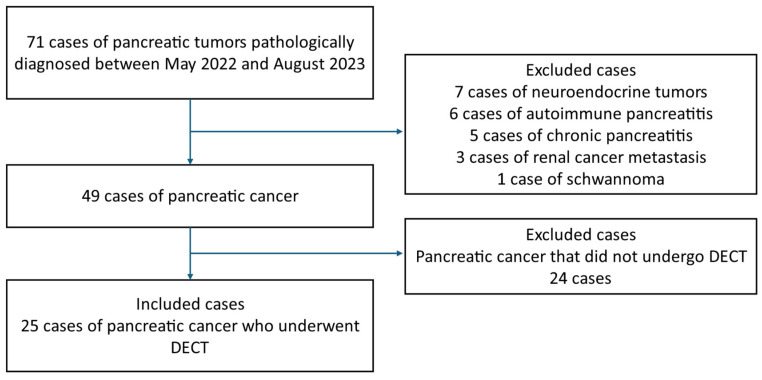
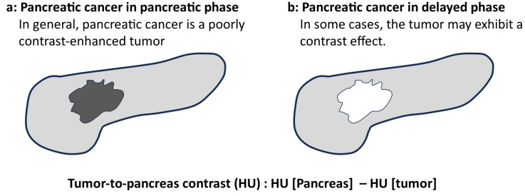
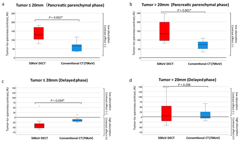
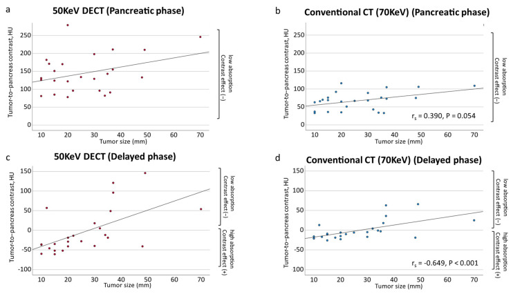
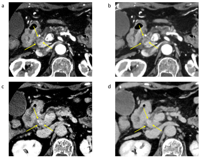
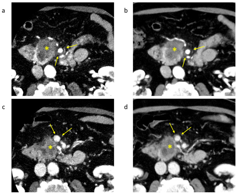
Background/Objectives: The usefulness of dual-energy computed tomography (DECT) for low absorption in the parenchymal phase and contrast effects in the delayed phase for pancreatic cancer is not clear. Therefore, the diagnostic capability of low-KeV images obtained using DECT for pancreatic cancer in the pancreatic parenchymal and delayed phases was evaluated quantitatively and qualitatively. Methods: Twenty-five patients with pancreatic cancer who underwent contrast-enhanced DECT were included. A total of 50 and 70 KeV CT images, classified as low-keV and conventional CT-equivalent images, were produced, respectively. The tumor-to-pancreas contrast (Hounsfield units [HU]) in the pancreatic parenchymal and delayed phases was calculated by subtracting the CT value of the pancreatic tumor from that of normal parenchyma. Results: The median tumor-to-pancreas contrast on 50 KeV CT in the pancreatic parenchymal phase (133 HU) was higher than that on conventional CT (68 HU) (p < 0.001). The median tumor-to-pancreas contrast in the delayed phase was -28 HU for 50 KeV CT and -9 HU for conventional CT (p = 0.545). For tumors < 20 mm, the tumor-to-pancreas contrast of 50 KeV CT (-39 HU) had a significantly clearer contrast effect than that of conventional CT (-16.5 HU), even in the delayed phase (p = 0.034). Conclusions: These 50 KeV CT images may clarify the low-absorption areas of pancreatic cancer in the pancreatic parenchymal phase. A good contrast effect was observed in small pancreatic cancers on 50 KeV delayed-phase images, suggesting that DECT is useful for the visualization of early pancreatic cancer with a small tumor diameter.

Keywords: contrast; delayed phase; dual-energy computed tomography; pancreatic cancer.


Conflict of interest statement

The authors declare no conflicts of interest.

Figures

Figure 1
Figure 1
The patient introduction flowchart.
Figure 2
Figure 2
The overview of the quantitative evaluation (created by the author). (a) Pancreatic cancer in the pancreatic phase. In general, pancreatic cancer is depicted as a hypoenhanced tumor on CT. (b) Pancreatic cancer in the delayed phase. In some cases, the tumor is depicted as a hyperenhanced tumor on CT.
Figure 3
Figure 3
Tumor-to-pancreas contrast (50 KeV DECT vs. conventional CT [70 KeV]). In the pancreatic parenchymal phase CT, the contrast between the tumor and surrounding pancreatic parenchyma was significantly larger at the 50 KeV setting in both ≤20 mm (a) and >20 mm lesions (b). In the delayed-phase CT, a tumor of ≤20 mm could be detected with significantly higher contrast between the tumor and surrounding pancreatic parenchyma at the 50 KeV rather than 70 KeV setting (c), but there was no significant difference in the contrast between 50 KeV and 70 KeV in the >20 mm lesion (d). * Statistically significant.
Figure 4
Figure 4
Scatter plots of the contrast difference between tumor and pancreatic parenchyma by tumor diameter. (a) Tumor-to-pancreas contrast on 50 KeV DECT in the pancreatic parenchymal phase (rs = 0.270, p = 0.192). (b) Tumor-to-pancreas contrast of conventional CT (70 KeV) in the pancreatic parenchymal phase (rs = 0.390, p = 0.054). (c) Tumor-to-pancreas contrast of 50 KeV DECT in the delayed phase (rs = −0.628, p < 0.001 *). (d) Tumor-to-pancreas contrast on conventional CT (70 KeV) in the delayed phase (rs = −0.649, p < 0.001 *). * Statistically significant.
Figure 5
Figure 5
Case of a 77-year-old woman with pancreatic cancer 13 mm in size (Yokohama City University). (a) A 50 KeV DECT image was obtained in the pancreatic parenchymal phase. (b) A conventional CT image (70 KeV image) was obtained in the pancreatic parenchymal phase. A low-absorption tumor is seen in the area indicated by the arrow. The contrast difference between the tumor and pancreas (tumor-to-pancreas contrast (HU): HU [pancreas] − HU [tumor]) is 171 HU in the 50 KeV DECT image and 70 HU in the conventional CT image (70 KeV), showing the tumor more clearly in the 50 KeV DECT image. (c) The 50 KeV DECT image was obtained in the delayed phase. (d) A conventional CT image (70 KeV image) was obtained in the delayed phase. The tumor-to-pancreas contrast was −46 HU for the 50 KeV image and −17 HU for the conventional CT image (70 KeV image), indicating that the 50 KeV DECT image clearly showed the contrast effect in the delayed phase.
Figure 6
Figure 6
Case of a 75-year-old man with pancreatic cancer (Yokohama City University). *: Tumor area of pancreatic cancer. (a) A 50 KeV DECT image was obtained in the pancreatic parenchymal phase. (b) A conventional CT image (70 KeV image) was obtained in the pancreatic parenchymal phase. The tip of the arrow is the superior mesenteric artery, which is in contact with the tumor; the 50 KeV DECT image more clearly suggests dorsal involvement of the superior mesenteric artery beyond 180°. (c) A 50 KeV DECT image was obtained in the portal phase. (d) A conventional image (70 KeV image) was obtained in the portal phase. The tip of the arrow is the superior mesenteric vein, which is in contact with the tumor.

Similar articles

References

    1. Siegel R.L., Miller K.D., Fuchs H.E., Jemal A. Cancer Statistics, 2021. CA A Cancer J. Clin. 2021;71:7–33. doi: 10.3322/caac.21654. - DOI - PubMed
    1. Siegel R., Ma J., Zou Z., Jemal A. Cancer statistics, 2014. CA A Cancer J. Clin. 2014;64:9–29. doi: 10.3322/caac.21208. - DOI - PubMed
    1. Vincent A., Herman J., Schulick R., Hruban R.H., Goggins M. Pancreatic cancer. Lancet. 2011;378:607–620. doi: 10.1016/S0140-6736(10)62307-0. - DOI - PMC - PubMed
    1. Ryan D.P., Hong T.S., Bardeesy N. Pancreatic adenocarcinoma. N. Engl. J. Med. 2014;371:1039–1049. doi: 10.1056/NEJMra1404198. - DOI - PubMed
    1. Blouhos K., Boulas K.A., Tsalis K., Hatzigeorgiadis A. The isoattenuating pancreatic adenocarcinoma: Review of the literature and critical analysis. Surg. Oncol. 2015;24:322–328. doi: 10.1016/j.suronc.2015.09.006. - DOI - PubMed