

Protective effects of Silibinin and cinnamic acid against paraquat-induced lung toxicity in rats: impact on oxidative stress, PI3K/AKT pathway, and miR-193a signaling
- PMID: 39453500
- PMCID: PMC11978700
- DOI: 10.1007/s00210-024-03511-y
Protective effects of Silibinin and cinnamic acid against paraquat-induced lung toxicity in rats: impact on oxidative stress, PI3K/AKT pathway, and miR-193a signaling
Abstract
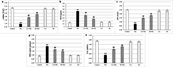
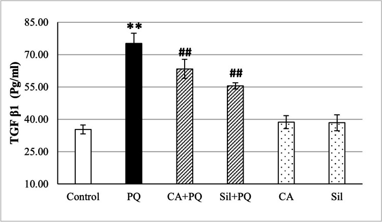
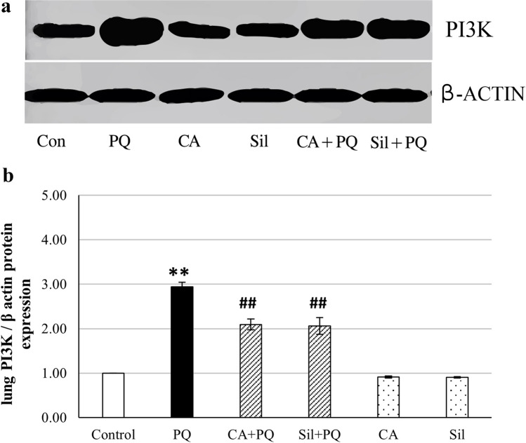
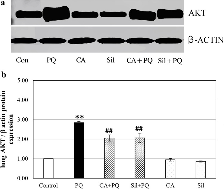
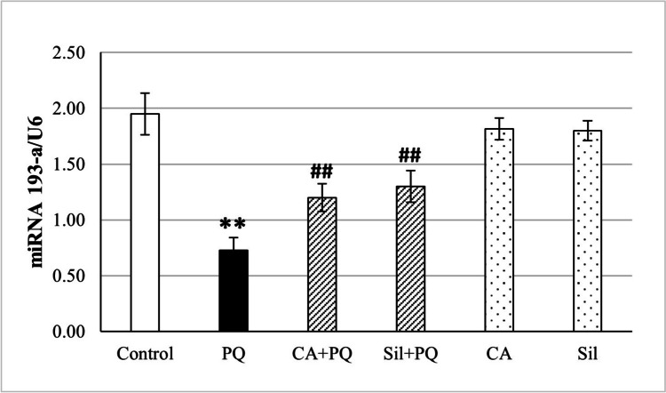
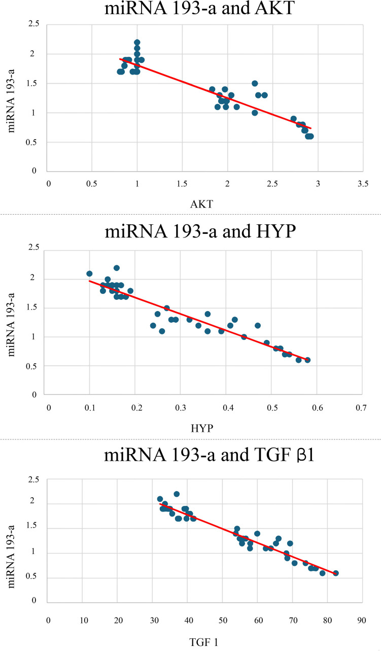
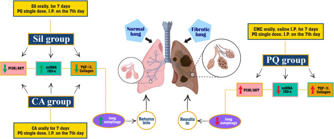
Levels of reactive oxygen species (ROS) are the primary determinants of pulmonary fibrosis. It was discovered that antioxidants can ameliorate pulmonary fibrosis caused by prolonged paraquat (PQ) exposure. However, research on the precise mechanisms by which antioxidants influence the signaling pathways implicated in pulmonary fibrosis induced by paraquat is still insufficient. This research utilized a rat model of pulmonary fibrosis induced by PQ to examine the impacts of Silibinin (Sil) and cinnamic acid (CA) on pulmonary fibrosis, with a specific focus on pro-fibrotic signaling pathways and ROS-related autophagy. Lung injury induced by paraquat was demonstrated to be associated with oxidative stress and inflammation of the lungs, downregulated (miR-193a), and upregulated PI3K/AKT/mTOR signaling lung tissues. Expression levels of miR-193a were determined with quantitative real-time PCR, protein level of protein kinase B (Akt), and phosphoinositide 3-Kinase (PI3K) which were determined by western blot analysis. Hydroxyproline levels (HYP) and transforming growth factor-β1 (TGF-β1) were measured by ELISA, malondialdehyde (MDA), total antioxidant capacity (TAC), glutathione peroxidase (GSH), and catalase and were measured in lung tissue homogenates colorimetrically using spectrophotometer. Long-term exposure to paraquat resulted in decreased PI3K/AKT signaling, decreased cell autophagy, increased oxidative stress, and increased pulmonary fibrosis formation. Silibinin and cinnamic acid also decreased oxidative stress by increasing autophagy and miR-193a expression, which in turn decreased pulmonary fibrosis. These effects were associated by low TGF-β1. Silibinin and cinnamic acid inhibited PQ-induced PI3K/AKT by stimulating miR-193-a expression, thus attenuating PQ-induced pulmonary fibrosis.
Keywords: AKT; Cinnamic acid; MiRNA 193a; PI3K; Paraquat; Silibinin.
© 2024. The Author(s).
Conflict of interest statement
Declarations. Ethical approval: This research was approved by BSU-IACUC reviewers and the approval number is (022-529). The National Institutes of Health’s (NIH) guide for the care and use of Laboratory animals (NIH Publications No. 8023, revised 1978) and the local institutional Research Ethics Committee’s approval were both followed in the carrying out of each experiment in respect to the ARRIVE guidelines (Animal Research: Reporting of In-Vivo Experiments). Consent to participate: Not applicable. Consent for publication: Not applicable. Conflict of interests: The authors declare no competing interests.
Figures
Similar articles
-
Ligustrazin increases lung cell autophagy and ameliorates paraquat-induced pulmonary fibrosis by inhibiting PI3K/Akt/mTOR and hedgehog signalling via increasing miR-193a expression.BMC Pulm Med. 2019 Feb 11;19(1):35. doi: 10.1186/s12890-019-0799-5. BMC Pulm Med. 2019. PMID: 30744607 Free PMC article.
-
Radix puerariae extracts ameliorate paraquat-induced pulmonary fibrosis by attenuating follistatin-like 1 and nuclear factor erythroid 2p45-related factor-2 signalling pathways through downregulation of miRNA-21 expression.BMC Complement Altern Med. 2016 Jan 12;16:11. doi: 10.1186/s12906-016-0991-6. BMC Complement Altern Med. 2016. PMID: 26758514 Free PMC article.
-
Amelioration of paraquat-induced pulmonary fibrosis in mice by regulating miR-140-5p expression with the fibrogenic inhibitor Xuebijing.Int J Immunopathol Pharmacol. 2020 Jan-Dec;34:2058738420923911. doi: 10.1177/2058738420923911. Int J Immunopathol Pharmacol. 2020. PMID: 32462952 Free PMC article.
-
Pulmonary toxicity and fibrogenic response of carbon nanotubes.Toxicol Mech Methods. 2013 Mar;23(3):196-206. doi: 10.3109/15376516.2012.753967. Epub 2013 Jan 16. Toxicol Mech Methods. 2013. PMID: 23194015 Free PMC article. Review.
-
A particle of concern: explored and proposed underlying mechanisms of microplastic-induced lung damage and pulmonary fibrosis.Inhal Toxicol. 2025 Jan;37(1):1-17. doi: 10.1080/08958378.2025.2461048. Epub 2025 Feb 11. Inhal Toxicol. 2025. PMID: 39932476 Review.
References
-
- Abd El-Raouf OM, El-Sayed ESM, Manie MF (2015) Cinnamic acid and cinnamaldehyde ameliorate cisplatin-induced splenotoxicity in rats. J Biochem Mol Toxicol 29:426–431 - PubMed
-
- Ahmed MA, El Morsy EM, Ahmed AA (2019) Protective effects of febuxostat against paraquat-induced lung toxicity in rats: impact on RAGE/PI3K/Akt pathway and downstream inflammatory cascades. Life Sci 221:56–64 - PubMed
-
- Barceloux DG (2009) Cinnamon (cinnamomum species). Disease-a-Month 55:327–335 - PubMed
-
- Chen Y-L, Huang S-T, Sun F-M, Chiang Y-L, Chiang C-J, Tsai C-M, Weng C-J (2011) Transformation of cinnamic acid from trans-to cis-form raises a notable bactericidal and synergistic activity against multiple-drug resistant Mycobacterium tuberculosis. Eur J Pharm Sci 43:188–194 - PubMed
MeSH terms
Substances
LinkOut - more resources
Full Text Sources
Medical
Miscellaneous
