. 2024 Oct 29;121(44):e2405806121.
doi: 10.1073/pnas.2405806121. Epub 2024 Oct 25.

Arabidopsis uses a molecular grounding mechanism and a biophysical circuit breaker to limit floral abscission signaling

Affiliations

Arabidopsis uses a molecular grounding mechanism and a biophysical circuit breaker to limit floral abscission signaling

Isaiah W Taylor et al. Proc Natl Acad Sci U S A. .

Abstract

Abscission is the programmed separation of plant organs. It is widespread in the plant kingdom with important functions in development and environmental response. In Arabidopsis, abscission of floral organs (sepals, petals, and stamens) is controlled by two receptor-like protein kinases HAESA (HAE) and HAESA LIKE-2 (HSL2), which orchestrate the programmed dissolution of the abscission zone connecting floral organs to the developing fruit. In this work, we use single-cell RNA sequencing to characterize the core HAE/HSL2 abscission gene expression program. We identify the MAP KINASE PHOSPHATASE-1/MKP1 gene as a negative regulator of this pathway. MKP1 acts prior to activation of HAE/HSL2 signaling to establish a signaling threshold required for the initiation of abscission. Furthermore, we use single-cell data to identify genes expressed in two subpopulations of abscission zone cells: those proximal and those distal to the plane of separation. We identify INFLORESCENCE DEFICIENT IN ABSCISSION/IDA family genes, encoding activating ligands of HAE/HSL2, as enriched in distal abscission zone cells at the base of the abscising organs. We show how this expression pattern forms a biophysical circuit breaker whereby, when the organ is shed, the source of the IDA peptides is removed, leading to cessation of HAE/HSL2 signaling. Overall, this work provides insight into the multiple control mechanisms acting on the abscission-signaling pathway.

Keywords: abscission; signaling; singe-cell rna-sequencing.


Conflict of interest statement

Competing interests statement:The authors declare no competing interest.

Figures
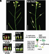
Fig. 1.
Fig. 1.
Background and experimental system. (A) Floral abscission phenotype of WT and hae hsl2 mutant. (Scale bar, 10 mm.) (B) Flower stages in WT and hae hsl2 mutant. (Scale bar, 2 mm.) (C) Phenotype of transgenic hae hsl2 expressing wild-type HAEpr::HAE-YFP and HAEpr::HAE-YFP K711E (bright-field/BF and YFP). (Scale bar, 2 mm.) (D) Diagram of AZ single-cell isolation and experimental procedure.
Fig. 2.
Fig. 2.
Identification and characterization of AZ cells by single-cell RNA sequencing. (A) UMAP embedding of WT cells with putative AZ cluster circles in pink (Top) and expression of AZ marker genes on UMAP (Bottom). Expression is on Seurat SCT scale. (B) Cluster-wise pseudobulk Spearman correlation with sorted bulk data of HAEpr::HAE-YFP (Top) or QRT2pr::GFP (Bottom). (C) Expression of 4 putative AZ marker genes in single-cell UMAP embedding and in the young siliques of promoter::fluorescent protein expressing transgenic plants (bright-field/BF and YFP or GFP). Expression is on Seurat SCT scale. (Scale bar, 0.7 mm.) (D) Gene Ontology Biological Process term enrichment of AZ-specific genes.
Fig. 3.
Fig. 3.
Analysis of differentially expressed genes in the hae hsl2 mutant AZ. (A) UMAP embedding of hae hsl2 cells. (B) Gene Ontology Biological Process term enrichment of DE genes higher in WT (FDR <.05, log2(FC) > 1). (C) Expression of cell wall/middle lamella hydrolysis genes (Top) and cutin/lignin biosynthesis genes (Bottom). Scale is log2(FC) for each sample relative to the average of hae hsl2. All genes are lower in mutants with FDR < 0.05.
Fig. 4.
Fig. 4.
Analysis and identification of hae hsl2 suppressors. (A) Phenotypes of WT, hae hsl2, hae hsl2 fal-3, and hae hsl2 fal-7 at 22° (Left) or 16° (Right). Siliques were gently tapped to remove remnant floral organs. (Scale bar, 1 mm.) (B) Breakstrength phenotypes of hae hsl2, hae hsl2 fal-3, and hae hsl2 fal-7 at 23° (Left) or 16° (Right). (C) Heatmap of expression values for 67 genes identified as higher in WT in both bulk and single-cell DE analysis comparing WT to hae hsl2. Values are log2(FC) compared to the overall average expression across all genotypes. (D) Average log2(FC) comparing each sample to the average of hae hsl2 for the genes in part C. (E) Complementation crosses of fal mutants. (Scale bar, 1 mm.) (F) Gene model of MKP1 depicting mutations in fal-3 and fal-7. Orange represents exons, blue represents UTRs, and thin lines represent introns.
Fig. 5.
Fig. 5.
Spatial analysis of the AZ. (A) Schematic view of the spatial organization of the AZ. (B) Low-resolution Louvain clustering and UMAP embedding of WT AZ cells (Left) and Spearman correlation with sorted bulk secession data (Middle) or residuum data (Right). (C) GO Term enrichment of genes differentially enriched in the putative secession cells (Top panel) or putative residuum cells (Bottom panel). (D) Pseudobulk log2(CPM + 1) of AT5G57785 in putative secession (“sec.”) and residuum (“res.”) cells. (E) AT5G57785pr::H2B-VENUS expression in secession and residuum cells. (F) Pseudobulk log2(CPM + 1) of IDA in putative secession and residuum cells. (G) IDApr::H2B-VENUS expression in secession and residuum cells. (H) IDA expression phenotype in WT (Left) and residuum-specific AT5G57785pr::IDA misexpression phenotype (Right).
Fig. 6.
Fig. 6.
Diagram depicting the molecular signaling circuit controlling abscission signal initiation and termination. References to the “IDA peptide signal” refer to peptides produced by the IDA and redundant IDA-LIKE genes.

Similar articles

Cited by

References

    1. Addicott F. T., Lynch R. S., Physiology of abscission. Ann. Rev. Plant Physiol. 6, 211–238 (1955).
    1. Taylor J. E., Whitelaw C. A., Signals in abscission. New Phytol. 151, 323–340 (2001).
    1. Patharkar O. R., Walker J. C., Connections between abscission, dehiscence, pathogen defense, drought tolerance, and senescence. Plant Sci. 284, 25–29 (2019). - PubMed
    1. Yu Y., Kellogg E. A., Inflorescence abscission zones in grasses: Diversity and genetic regulation. Ann. Plant Rev. 1, 1–35 (2018).
    1. Dutta S. K., Gurung G., Yadav A., Laha R., Mishra V. K., Factors associated with citrus fruit abscission and management strategies developed so far: A review. N Zeal. J. Crop Horticultural Sci. 51, 467–488 (2022).

MeSH terms

Substances

LinkOut - more resources