. 2024 Dec 1;110(12):7563-7572.
doi: 10.1097/JS9.0000000000002128.

Low-energy extracorporeal shockwave therapy improves locomotor functions, tissue regeneration, and modulating the inflammation induced FGF1 and FGF2 signaling to protect damaged tissue in spinal cord injury of rat model: an experimental animal study

Affiliations

Low-energy extracorporeal shockwave therapy improves locomotor functions, tissue regeneration, and modulating the inflammation induced FGF1 and FGF2 signaling to protect damaged tissue in spinal cord injury of rat model: an experimental animal study

Chieh-Cheng Hsu et al. Int J Surg. .

Abstract

Background: Spinal cord injury (SCI) is a debilitating condition that results in severe motor function impairments. Current therapeutic options remain limited, underscoring the need for novel treatments. Extracorporeal shockwave therapy (ESWT) has emerged as a promising noninvasive approach for treating musculoskeletal disorders and nerve regeneration.

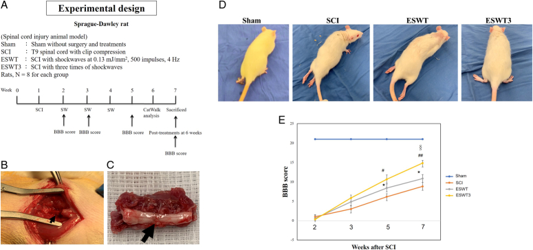
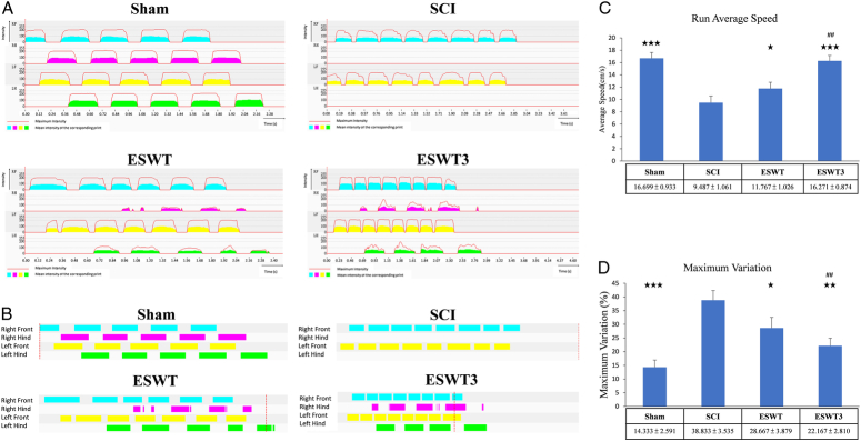
Methods: This study explored the effects of low-energy ESWT on locomotor function, tissue regeneration, inflammation, and mitochondrial function in a rat SCI model. Experiments were performed using locomotor function assays, CatWalk gait analysis, histopathological examination, immunohistochemical, and immunofluorescence staining.

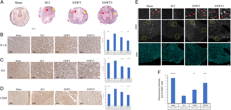
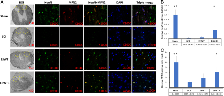
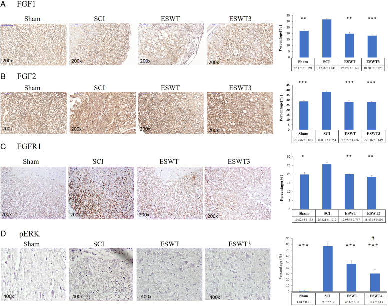
Results: The findings demonstrated that low-energy ESWT had a dose-dependent effect, with three treatment sessions (ESWT3) showing superior outcomes compared to a single session. ESWT3 significantly improved motor functions [run patterns, run average speed, and maximum variation, as well as the Basso, Beattie, and Bresnahan score] and promoted tissue regeneration while reducing inflammation. ESWT3 significantly decreased levels of IL-1β, IL6, and macrophages (CD68) while increasing leukocyte (CD45) infiltration. Additionally, ESWT3 upregulated NueN and mitofusin 2 (MFN2), suggesting enhanced neuronal health and mitochondrial function. Moreover, ESWT3 modulated the expression of fibroblast growth factor 1 (FGF1), FGF2, their receptor FGFR1 and phosphorylation of ERK, aiding tissue repair, and regeneration in SCI.

Conclusions: This study highlights the potential of low-energy ESWT as an effective noninvasive treatment for SCI, demonstrating significant improvements in motor recovery, tissue regeneration, anti-inflammatory effects, and mitochondrial protection. These findings provide valuable insights into the mechanisms of ESWT and its therapeutic application for SCI recovery.


Conflict of interest statement

The authors declare no conflicts of interest.

Sponsorships or competing interests that may be relevant to content are disclosed at the end of this article.

Figures

Figure 1
Figure 1
The study design and locomotor function assay. (A) The graph presents the study design of the experiment including Sham, SCI, ESWT, and ESWT3 groups. ESWT is that shockwaves applied at 1 week postsurgery and ESWT3 is that shockwaves applied at 1, 2, and 3 weeks postsurgery, respectively. The sacrifice of animals are at 6 weeks postsurgery. Eight rats are used for each group. (B) The injury is created at T9 of the spinal cord in the rat as indicated by the black arrow. (C) The damage region of the spinal cord is indicated by the black arrow. (D) The hind legs of the rat drag in the SCI and ESWT groups. Some of the rats have their paws turned upwards in the ESWT3 group at 6 weeks postsurgery. (E) The BBB scores are presented in the Sham, SCI, ESWT, and ESWT3 groups at 2, 3, 5, and 7 weeks. * P<0.05 compared between ESWT and SCI groups. # P<0.05 and ## P<0.01 are compared between ESWT3 and SCI group. § P<0.05 compared between ESWT and ESWT3 group. BBB, Basso, Beattie, and Bresnahan; ESWT, extracorporeal shockwave therapy; ESWT3, three ESWT treatments; N=8; SCI, spinal cord injury.
Figure 2
Figure 2
The graphical print view and data for each group by gait analysis from CatWalk. (A) The intensity of the run patterns. (B) Print view of the run pattern. (C) Run average speed. (D) Maximum variation. The *** P<0.001, ** P<0.01, and * P<0.05 are compared with the SCI group. The ## P<0.01 are compared between ESWT and ESWT3 groups. N=8. ESWT, extracorporeal shockwave therapy; ESWT3, three ESWT treatments; SCI, spinal cord injury.
Figure 3
Figure 3
The histological structure, inflammation cytokines and immunocells measurement in the spinal cord injury after treatments. (A) The morphological observation is performed in the Sham, SCI, ESWT, and ESWT3 groups. The scale bar is 500 μm. The red arrow is indicated gray matter and yellow arrow is indicated white matter in the spinal cord. (B) The immunohistochemistry staining of IL1-β in the Sham, SCI, ESWT, and ESWT groups. The right panel is the expressed percentage of IL1-β. (C) The immunohistochemistry staining of IL6 in the Sham, SCI, ESWT, and ESWT groups. The right panel is the expressed percentage of IL6. The images are 200x magnification. (D) The immunohistochemistry staining of CD68-positive macrophages in the Sham, SCI, ESWT, and ESWT groups. The right panel is the expressed percentage of CD68-positive macrophages. The images are 200x magnification scale bar is 100 μm. (E) The immunofluorescence staining of CD45-positive leukocyte in the Sham, SCI, ESWT, and ESWT groups. Red arrows: increased leukocyte infiltration. Scale bar: 100 μm. (F) The panel is the fluorescence intensity of CD45-positive leukocyte. The *** P<0.001, ** P<0.01, and * P<0.05 are compared with the SCI group. N=4. ESWT, extracorporeal shockwave therapy; ESWT3, three ESWT treatments; SCI, spinal cord injury.
Figure 4
Figure 4
The NeuN and MFN2 measurements in the spinal cord injury after treatments. (A) The immunofluorescence staining of NeuN and MFN2 in the Sham, SCI, ESWT, and ESWT3 groups. (B) The related NeuN positive cells. (C) The related MFN2 positive cells. The ** P<0.01 and * P<0.05 are compared with SCI group. N=8. ESWT, extracorporeal shockwave therapy; ESWT3, three ESWT treatments; ROI, region of interest; SCI, spinal cord injury. The bright field of the images is 50x magnification. The images of immunofluorescence staining is 1000x magnification.
Figure 5
Figure 5
The expression of inflammation induced FGF1, FGF2, and FGFR1 receptor in the spinal cord injury after treatments. (A) The immunohistochemistry staining of FGF1-positive cells in the Sham, SCI, ESWT, and ESWT groups. The right panel is the expressed percentage of FGF1-positive cells. The images are 200x magnification. (B) The immunohistochemistry staining of FGF2-positive cells in the Sham, SCI, ESWT, and ESWT groups. The right panel is the expressed percentage of FGF2-positive cells. The images are 200x magnification. (C) The immunohistochemistry staining of FGFR1-positive cells in the Sham, SCI, ESWT, and ESWT groups. The right panel is the expressed percentage of FGFR1-positive cells. The images are 200x magnification. (D) The immunohistochemistry staining of pERK-positive cells in the Sham, SCI, ESWT, and ESWT groups. The right panel is the expressed percentage of pERK-positive cells. The *** P<0.001, ** P<0.01, and * P<0.05 are compared with the SCI group. # P<0.05 is compared with the ESWT group. N=8. ESWT, extracorporeal shockwave therapy; ESWT3, three ESWT treatments; SCI, spinal cord injury.

References

    1. Wang TY, Park C, Zhang H, et al. . Management of acute traumatic spinal cord injury: a review of the literature. Front Surg 2021;8:698736. - PMC - PubMed
    1. Li S, Dinh HTP, Matsuyama Y, et al. . Molecular mechanisms in the vascular and nervous systems following traumatic spinal cord injury. Life 2022;13:9. - PMC - PubMed
    1. Zhang Y, Al Mamun A, Yuan Y, et al. . Acute spinal cord injury: pathophysiology and pharmacological intervention (Review). Mol Med Rep 2021;23:417. - PMC - PubMed
    1. Hellenbrand DJ, Quinn CM, Piper ZJ, et al. . Inflammation after spinal cord injury: a review of the critical timeline of signaling cues and cellular infiltration. J Neuroinflammation 2021;18:284. - PMC - PubMed
    1. Jaerve A, Muller HW. Chemokines in CNS injury and repair. Cell Tissue Res 2012;349:229–248. - PubMed

Substances