. 2024 Oct 25;10(43):eadp1053.
doi: 10.1126/sciadv.adp1053. Epub 2024 Oct 25.

Structural basis for regulation of a CBASS-CRISPR-Cas defense island by a transmembrane anti-σ factor and its ECF σ partner

Affiliations

Structural basis for regulation of a CBASS-CRISPR-Cas defense island by a transmembrane anti-σ factor and its ECF σ partner

Diego Bernal-Bernal et al. Sci Adv. .

Abstract

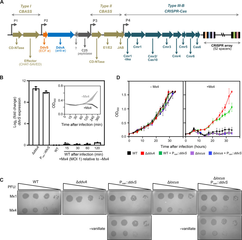
How CRISPR-Cas and cyclic oligonucleotide-based antiphage signaling systems (CBASS) are coordinately deployed against invaders remains unclear. We show that a locus containing two CBASS and one type III-B CRISPR-Cas system, regulated by the transmembrane anti-σ DdvA and its cognate extracytoplasmic function (ECF) σ DdvS, can defend Myxococcus xanthus against a phage. Cryo-electron microscopy reveals DdvA-DdvS pairs assemble as arrow-shaped transmembrane dimers. Each DdvA periplasmic domain adopts a separase/craspase-type tetratricopeptide repeat (TPR)-caspase HetF-associated with TPR (TPR-CHAT) architecture with an incomplete His-Cys active site, lacking three α-helices conserved among CHAT domains. Each active site faces the dimer interface, raising the possibility that signal-induced caspase-like DdvA autoproteolysis in trans precedes RseP-mediated intramembrane proteolysis and DdvS release. Nuclear magnetic resonance reveals a DdvA cytoplasmic CHCC-type zinc-bound three-helix bundle that binds to DdvS σ2 and σ4 domains, undergoing σ4-induced helix extension to trap DdvS. Altogether, we provide structural-mechanistic insights into membrane anti-σ-ECF σ regulation of an antiviral CBASS-CRISPR-Cas defense island.


Figures

Fig. 1.
Fig. 1.. M. xanthus DdvA-DdvS–dependent regulon and role in defense against myxophages.
(A) Schematic of the M. xanthus defense island regulated by DdvA-DdvS. DdvS-dependent promoters P1 to P4 (23) drive expression of genes encoding (left to right): (i) predicted type I CBASS with a CD-Ntase and an effector comprising N-terminal CHAT and a C-terminal cyclic oligonucleotide-sensing SAVED (SMODS-associated and fused to various effector domains, where SMODS is second messenger oligo- or dinucleotide synthetase domain) modules; (ii) DdvS, DdvA, a protein of unknown function, and a putative C25-type peptidase; (iii) type II CBASS with CD-NTase, ubiquitin transferase-like (E1/E2), and deubiquitinase-like (JAB) proteins; (iv) type III-B Cmr proteins and CRISPR array. (B) Analysis of ddvS expression by quantitative reverse transcription PCR (qRT-PCR) (means ± SEs, n = 3 biological replicates) in the WT strain, the ΔddvA strain, WT with vanillate-induced (0.5 mM vanillate) ddvS expression from promoter Pvan at a heterologous site, or WT infected or not with Mx4 at an MOI of 1 for the indicated post-infection times (inset, OD550 of cultures after infection with Mx4 at MOI = 1 and the uninfected equivalent). (C) Plaque assays performed at 26°C with Mx1 and Mx4 spotted in 10-fold serial dilutions (PFU, plaque-forming units) on a lawn of the indicated strains: WT, ΔddvA, WT Pvan::ddvS, Δlocus, Δlocus Pvan::ddvS. For strains with Pvan::ddvS, growth media with 0.5 mM vanillate (top) or without (bottom) were used. (D) Growth curves for the strains in (C) (0.5 mM vanillate present for strains with Pvan::ddvS) with or without Mx4 infection (0.1 MOI).
Fig. 2.
Fig. 2.. Architecture of the DdvA dimer in complex with DdvS.
(A) Two views of the structure of the transmembrane anti-σ DdvA dimer in complex with its cognate ECF σ DdvS determined by cryo-EM with that of the cytoplasmic region from NMR. Schematic of the primary structure of the DdvA dimer below (NTD corresponds to DdvANt and the scaffold-TPR-CHAT region corresponds to DdvACt) with the color code for the different regions used in all panels. (B) Cryo-EM density map at 2.94-Å resolution of the periplasmic DdvACt domain (residues 93 to 991) arranged as a dimer with an arrowhead-shaped architecture. (C) Cartoon representation of the DdvACt structure in (B). The inset (top left corner) shows a colored sketch to help navigate the panel. (D) Two views of the protomer structure of the DdvACt dimer with the helices (α) and strands (β) numbered and labeled, and domains colored as in (B).
Fig. 3.
Fig. 3.. DdvA CHAT catalytic dyad and the flexible linker connecting the TPR-CHAT domain and the TM helix.
(A) Overlay of the DdvA CHAT module (maroon) with that of the Desulfonema ishimotonii craspase in the inactive (left, pale blue; PDB 8EEX) and active (right, turquoise; PDB 8EEY) forms. A three α-helix motif (lime green) present in craspase is absent in DdvA. The catalytic dyad His and Cys side chains are represented as spheres (top) or as sticks in the zoom below with distances between their backbone Cα indicated. The His Nπ to Cys thiol S distances are 11.3 Å in DdvA, and 8.3 and 4.8 Å in inactive and active craspase, respectively. (B) Close-up of the DdvACt CHAT catalytic center of one protomer in the vicinity of a flexible sequence linking the TM helix and helix α5 of the opposite protomer. The periplasmic side of the membrane is depicted as above a line. In chain A, the flexible linker region (Q95-E102) following the TM helix is colored in black, the region closest to the catalytic dyad (V129-P140) is in cyan, and the rest of the linker is in orange. In chain B, the CHAT catalytic dyad residues H862 and C907 are shown as spheres. N-terminal fragments of DdvA truncated at Q128 and P140 were tested for RseP-dependent degradation. Bottom right, schematic with color codes for the DdvA regions.
Fig. 4.
Fig. 4.. CHAT catalytic sites in DdvACt, separase, and craspase, and RsePMx targeting of DdvA lacking its TPR-CHAT domain in vivo.
(A) DdvACt. (B) Yeast separase-securin complex (PDB ID: 5U1T). (C) D. ishimotonii active craspase (PDB ID: 8EEY). (D) Candidatus “Scalindua brodae” active craspase (PDB ID: 8EEY). Schemes below show color codes for distinct segments (numbered and labeled) along the polypeptide chain. CHAT domains are in maroon, TPR domains in blue, and the three α-helix motif in separase and craspase active sites absent in DdvA in lime green. In (A), chain B TPR-CHAT faces chain A midrib (light yellow). In (B) to (D), other domains are in light gray and subunits in dark gray; in (C), a retained craspase cleavage product is in purple. Catalytic dyad cysteine and histidine residues are shown as spheres and separase-bound inhibitor securin in stick representation. (E) Immunoblot of cell extracts of WT and ΔddvA strains expressing N-terminal FLAG-tagged DdvANt (DdvA1–70), DdvA1–140, and DdvA1–128 (or DdvA, as control) from an IPTG-inducible promoter (PIPTG) with 1 mM IPTG present. (F) Immunoblots of cell extracts from the ΔrsePMx strain (left) and its derivative with rsePMx controlled by a vanillate-inducible promoter (Pvan) for conditional expression (±0.5 mM vanillate present) at a heterologous site (right) expressing FLAG-tagged DdvANt, DdvA1–140, and DdvA1–128 as in (E). FLAG-tagged proteins were probed using anti-FLAG antibodies and the constitutively expressed CdnL protein (loading control below) with polyclonal anti-CdnL antibodies.
Fig. 5.
Fig. 5.. NMR solution structure of DdvANt and its interactions with DdvS.
(A) Average of the 20 final NMR structures of the three α-helix bundle DdvANt indicating α1, α2, α3, CHCC motif side chains as sticks (C, green; H, brown) and bound zinc (red sphere). H56 in the disordered C-terminal tail is represented by various conformations. (B) Superposition of the DdvANt NMR structure (dark blue) with RslA, RsiW, and ChrR ZASDs, or RseA and MucA non-zinc ASDs (PDB IDs: 3HUG, 5WUQ, 2Z2S, 1OR7, and 6IN7, respectively). (C) CSP (top), TALOS+ CSI (middle), and heteronuclear 15N{1H} NOEs (bottom) for each residue in [13C,15N]H6-DdvANt when free (dark blue), σ2S-bound (blue), or σ4S-bound (light blue). CSP cutoffs (dotted lines) are 0.15 for DdvANt-σ2S and 0.2 for DdvANt-σ4S. (D) Superposition of DdvANt free (dark blue) and σ4S-bound (light blue) NMR structures. Note a more fixed DdvANt H56 in a rigid α3 extension when σ4S-bound but unstructured in a flexible C-terminal tail when free or σ2S-bound.
Fig. 6.
Fig. 6.. Model of the DdvANt-DdvS complex and correlation with NMR data.
(A) Two views of the DdvANt-DdvS complex AlphaFold2 model colored. (B) Complex of DdvANt (blue, ribbon representation) with DdvS (σ2S, light gray; σ4S, dark gray; surface representation) with NMR CSP data indicated for σ2S-bound DdvANt (top) and σ4S-bound DdvANt (bottom). DdvANt CSPs >0.2 are primarily in the three-helix bundle with σ2S and in the α3 extension and C-terminal tail with σ4S. “N” and “C” correspond to the N and C termini of each protein and are colored accordingly.
Fig. 7.
Fig. 7.. Mutational analysis of DdvA-DdvS interactions.
(A) BACTH analysis of the interaction between DdvANt variants and full-length DdvS, σ2S, or σ4S domains on 40 μg ml−1 X-Gal/LB plates (blue colony color from reporter lacZ expression indicates interaction). C− is a representative negative control using cells with pKT25-DdvANt and empty pUT18C. (B) Plaque assays with myxophage Mx4 spotted in 10-fold serial dilutions on a lawn of the ΔddvA strain and its derivatives expressing C-terminally FLAG-tagged DdvA (“WT”) or its indicated variants from PIPTG with 0.1 mM IPTG present (left), aligned with qRT-PCR analysis of ddvS expression (means ± SEs, n = 3 biological replicates, data normalized relative to DdvA-FLAG WT) in these strains without Mx4 infection (right). (C) Immunoblots of cell extracts used in the qRT-PCR analysis in (B) with FLAG-tagged fusions probed using anti-FLAG antibodies and constitutively expressed CdnL protein (loading control) using polyclonal anti-CdnL antibodies.
Fig. 8.
Fig. 8.. Model for a proposed RIP mechanism in DdvA-DdvS signal transduction.
In the quiescent pre-S1P state with no signal, the bulky dimeric DdvA periplasmic TPR domain prevents the RseP intramembrane zinc protease (zinc, gray sphere) action on DdvA in complex with DdvS (top left) through steric hindrance of the tandem RseP PDZ domains. Signal-induced conformational changes activate DdvA CHAT protease activity for cleavage at the flexible linker that connects the TM helix to the bulky periplasmic TPR-CHAT domain to clip off the latter (top middle). This enables RseP-mediated intramembrane proteolysis of DdvA lacking its TPR-CHAT domain, release of the DdvA cytoplasmic domain bound to DdvS, followed by its dissociation and then degradation of the DdvA cytoplasmic domain by cytosolic proteases to liberate DdvS (top right). This leads to RNA polymerase–DdvS holoenzyme assembly and expression of the CBASS-CRISPR-Cas defense island and antiviral defense (bottom).

Similar articles

Cited by

  • Milestones in the development of Myxococcus xanthus as a model multicellular bacterium.
    Kroos L, Wall D, Islam ST, Whitworth DE, Muñoz-Dorado J, Higgs PI, Singer M, Mauriello EM, Treuner-Lange A, Søgaard-Andersen L, Kaimer C, Elías-Arnanz M, Stojković EA, Müller R, Volz C, Velicer GJ, Nan B. Kroos L, et al. J Bacteriol. 2025 Jul 24;207(7):e0007125. doi: 10.1128/jb.00071-25. Epub 2025 Jun 17. J Bacteriol. 2025. PMID: 40525847 Free PMC article. Review.
  • 'Intelligent' proteins.
    Tripathi T, Uversky VN, Giuliani A. Tripathi T, et al. Cell Mol Life Sci. 2025 Jun 14;82(1):239. doi: 10.1007/s00018-025-05770-1. Cell Mol Life Sci. 2025. PMID: 40515853 Free PMC article. Review.

References

    1. Makarova K. S., Wolf Y. I., Snir S., Koonin E. V., Defense islands in bacterial and archaeal genomes and prediction of novel defense systems. J. Bacteriol. 193, 6039–6056 (2011). - PMC - PubMed
    1. Doron S., Melamed S., Ofir G., Leavitt A., Lopatina A., Keren M., Amitai G., Sorek R., Systematic discovery of antiphage defense systems in the microbial pangenome. Science 359, eaar4120 (2018). - PMC - PubMed
    1. Millman A., Melamed S., Amitai G., Sorek R., Diversity and classification of cyclic-oligonucleotide-based anti-phage signalling systems. Nat. Microbiol. 5, 1608–1615 (2020). - PMC - PubMed
    1. Gao L., Altae-Tran H., Böhning F., Makarova K. S., Segel M., Schmid-Burgk J. L., Koob J., Wolf Y. I., Koonin E. V., Zhang F., Diverse enzymatic activities mediate antiviral immunity in prokaryotes. Science 369, 1077–1084 (2020). - PMC - PubMed
    1. Millman A., Melamed S., Leavitt A., Doron S., Bernheim A., Hör J., Garb J., Bechon N., Brandis A., Lopatina A., Ofir G., Hochhauser D., Stokar-Avihail A., Tal N., Sharir S., Voichek M., Erez Z., Ferrer J. L. M., Dar D., Kacen A., Amitai G., Sorek R., An expanded arsenal of immune systems that protect bacteria from phages. Cell Host Microbe 30, 1556–1569.e5 (2022). - PubMed

LinkOut - more resources