. 2024 Nov:77:103406.
doi: 10.1016/j.redox.2024.103406. Epub 2024 Oct 22.

Isoliquiritigenin alleviates cerebral ischemia-reperfusion injury by reducing oxidative stress and ameliorating mitochondrial dysfunction via activating the Nrf2 pathway

Affiliations

Isoliquiritigenin alleviates cerebral ischemia-reperfusion injury by reducing oxidative stress and ameliorating mitochondrial dysfunction via activating the Nrf2 pathway

Xiaobing Lan et al. Redox Biol. 2024 Nov.

Abstract

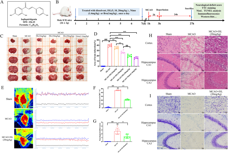
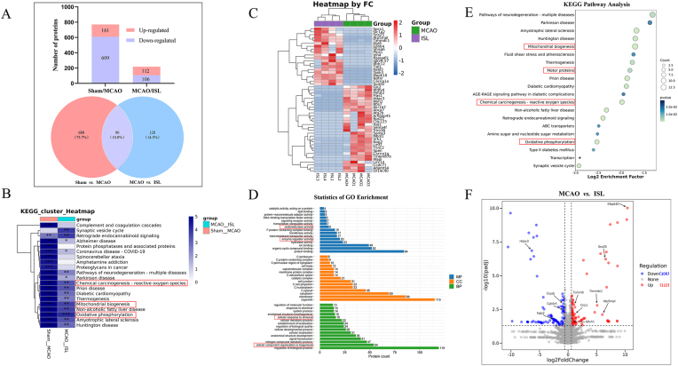
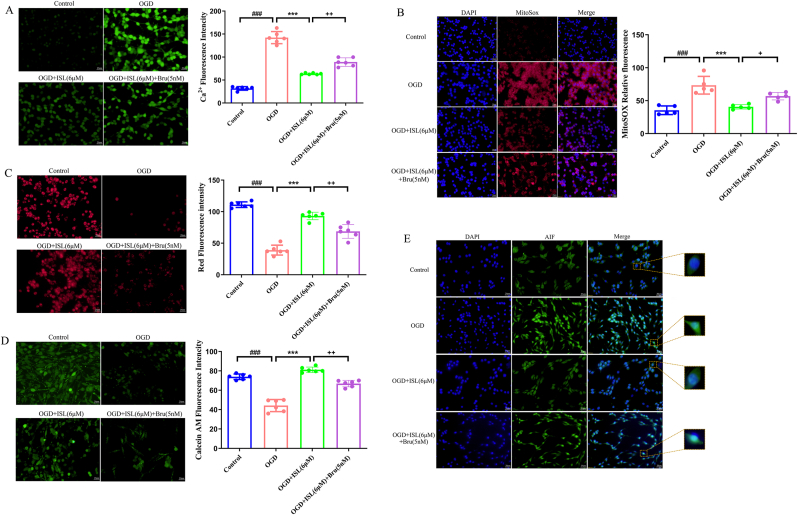
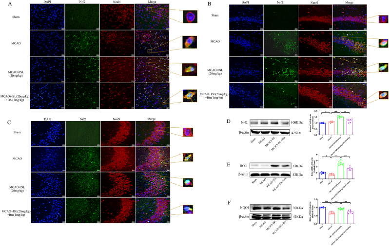
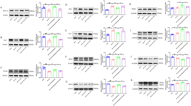
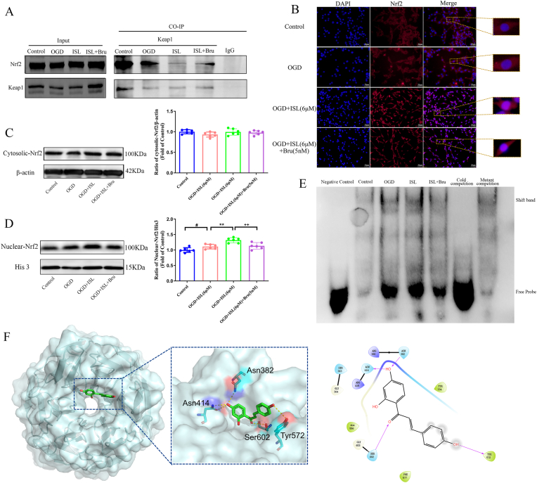
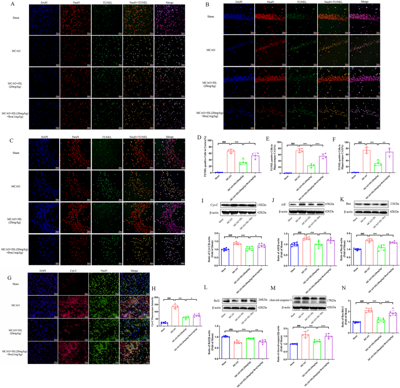
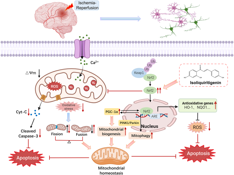
Cerebral ischemia-reperfusion injury (CIRI) refers to a secondary brain injury that occurs when blood supply is restored to ischemic brain tissue and is one of the leading causes of adult disability and mortality. Multiple pathological mechanisms are involved in the progression of CIRI, including neuronal oxidative stress and mitochondrial dysfunction. Isoliquiritigenin (ISL) has been preliminarily reported to have potential neuroprotective effects on rats subjected to cerebral ischemic insult. However, the protective mechanisms of ISL have not been elucidated. This study aims to further investigate the effects of ISL-mediated neuroprotection and elucidate the underlying molecular mechanism. The findings indicate that ISL treatment significantly alleviated middle cerebral artery occlusion (MCAO)-induced cerebral infarction, neurological deficits, histopathological damage, and neuronal apoptosis in mice. In vitro, ISL effectively mitigated the reduction of cell viability, Na+-K+-ATPase, and MnSOD activities, as well as the degree of DNA damage induced by oxygen-glucose deprivation (OGD) injury in PC12 cells. Mechanistic studies revealed that administration of ISL evidently improved redox homeostasis and restored mitochondrial function via inhibiting oxidative stress injury and ameliorating mitochondrial biogenesis, mitochondrial fusion-fission balance, and mitophagy. Moreover, ISL facilitated the dissociation of Keap1/Nrf2, enhanced the nuclear transfer of Nrf2, and promoted the binding activity of Nrf2 with ARE. Finally, ISL obviously inhibited neuronal apoptosis by activating the Nrf2 pathway and ameliorating mitochondrial dysfunction in mice. Nevertheless, Nrf2 inhibitor brusatol reversed the mitochondrial protective properties and anti-apoptotic effects of ISL both in vivo and in vitro. Overall, our findings revealed that ISL exhibited a profound neuroprotective effect on mice following CIRI insult by reducing oxidative stress and ameliorating mitochondrial dysfunction, which was closely related to the activation of the Nrf2 pathway.

Keywords: Cerebral ischemia-reperfusion injury; Isoliquiritigenin; Mitochondrial dysfunction; Neuroprotection; Nrf2 pathway.


Conflict of interest statement

Declaration of competing interest The authors declare that they have no conflict of interest.

Figures

Fig. 1
Fig. 1
ISL alleviated MCAO-induced brain injury, neurological deficit, and histopathological damage in mice. (A) The chemical information of ISL. (B) Flow chart of the experimental design. (C) Representative TTC staining images of coronal brain sections (the sections were presented “rostral to caudal”) at 24h after CIRI. (D) Quantitative analysis of brain infarct volume. (E) Monitoring images of CBF. (F) Quantitative analysis of the reduced rate of CBF in the ischemic brains. (G) Neurological deficit scores at 24 h after CIRI. (H and I) Representative HE and Nissl staining images of the histopathological changes in the ischemic cerebral cortex ( × 400, scale bar = 20 μm), hippocampus CA1 ( × 400, scale bar = 20 μm), and hippocampus CA3 ( × 200, scale bar = 50 μm) regions. Neurologic deficit scores are expressed as median with interquartile range, and normally distributed data are presented as the mean ± SD (n = 6). ###P < 0.001 versus sham group; ∗∗P < 0.01, ∗∗∗P < 0.001 versus MCAO group; ++P < 0.01 versus MCAO + ISL (5 mg/kg) group; &&P < 0.01 versus MCAO + ISL (10 mg/kg) group.
Fig. 2
Fig. 2
ISL prevented OGD-induced cell death and the consumption of antioxidase activities and alleviated DNA damage in PC12 cells. (A) Cell viability assay (n = 6). (B) Na+-K+-ATPase activity assay (n = 3). (C) MnSOD activity assay (n = 3). (D) Representative fluorescence images of comet assay in evaluating the degree of DNA damage, with a magnification of 400 × , scale bar = 20 μm. (E–J) Quantitative analysis of comet tail area, tail DNA, tail DNA (%), tail length, tail moment, and olive tail moment, respectively (n = 10). All values are expressed as the mean ± SD. ###P < 0.001 versus control group; ∗∗P < 0.01, ∗∗∗P < 0.001 versus OGD group.
Fig. 3
Fig. 3
DIA-Based quantitative proteomic analysis results (n = 4). (A) Statistical chart and Venn diagram of different protein changes in the sham vs. MCAO and MCAO vs. ISL groups. (B) Cluster analysis of the differential expressed proteins in the sham vs. MCAO and MCAO vs. ISL groups. (C) Heatmap of the top 50 differential expressed proteins in the MCAO and ISL group. (D) GO enrichment of those commonly selected differential proteins in the MCAO and ISL group, including the molecular function (blue), cellular component (orange), and biological process (green). (E) Bubble diagram of KEGG pathway analysis. (F) Volcano map of the differential expressed proteins in the MCAO and ISL group. All data values of |log2(Fold Change)| >0.585, Q-value <0.05.
Fig. 4
Fig. 4
ISL alleviated the oxidative stress damage and ameliorated the mitochondrial function after OGD injury in PC12 cells. (A) Representative F03 staining images of Ca2+ content in PC12 cells and statistical histogram, green fluorescence indicates the intensity of intracellular calcium ion fluorescence. (B) Representative MitoSOX staining images of mitochondrial ROS and statistical histogram, MitoSOX (red) represents the ROS contents in mitochondria, and DAPI (blue) was used to label the nuclei. (C) Representative TMRE staining images of MMP and statistical histogram, and red represents the fluorescence intensity of the TMRE cationic probe. (D) Representative Calcein AM staining images of MPTP and statistical histogram, and green represents the fluorescence intensity of the Calcein AM probe. (E) Representative immunofluorescent-stained images of AIF in the PC12 cells at 24 h after OGD injury, AIF (green) represents the expression of AIF-positive cells, and DAPI (blue) was used to label the nuclei. All the representative images are presented with a magnification of 400 × , scale bar = 20 μm. Data are expressed as the mean ± SD (n = 6). ###P < 0.001 versus control group; ∗∗∗P < 0.001 versus OGD group; +P < 0.05, ++P < 0.01 versus OGD + ISL group.
Fig. 5
Fig. 5
ISL treatment promoted the nuclear translocation of Nrf2 and the downstream antioxidant enzyme protein expressions at 24 h following ischemic insult in mice. Representative fluorescence images showed the expression of Nrf2 immuno-positive cells and nuclear translocation in the ischemic cerebral cortex (A), hippocampus CA1 (B), and hippocampus CA3 (C) regions after CIRI, with a magnification of 400 × , scale bar = 20 μm. Nrf2 (green) represents the expression of Nrf2-positive cells, NeuN (red) was used to label the neuron, and DAPI (blue) was used to label the nuclei. Representative Western blotting bands and quantification results of Nrf2 (D), HO-1 (E), and NQO1 (F) protein expression in ischemic brain tissues. Data are presented as the mean ± SD (n = 6). #P < 0.05, ###P < 0.001 versus sham group; ∗∗∗P < 0.001 versus MCAO group; ++P < 0.01, +++P < 0.001 versus MCAO + ISL group.
Fig. 6
Fig. 6
ISL improved the mitochondrial biogenesis, mitochondrial fusion-fission balance, and mitophagy. Representative Western blotting bands and quantitative analyses depicted the PGC1α (A), Nrf1 (B), TFAM (C), Drp1 (D), Fis1 (E), OPA1 (F), MFN2 (G), PINK1 (H), Parkin (I), p62 (J), LC3 (K) protein expression. Data are presented as the mean ± SD (n = 6). #P < 0.05, ##P < 0.01, ###P < 0.001 versus sham group; ∗∗P < 0.01, ∗∗∗P < 0.001 versus MCAO group; +P < 0.05, ++P < 0.01, +++P < 0.001 versus MCAO + ISL group.
Fig. 7
Fig. 7
ISL promoted the dissociation of Keap1/Nrf2 and the binding of Nrf2 with ARE in PC12 cells. (A) Co-IP assay showing the formation of Keap1/Nrf2 complex in each group. (B) Representative immunofluorescent-stained images of Nrf2 in PC12 cells following OGD injury at a magnification of 400 × , scale bar = 20 μm. Nrf2 (red) represents the expression of Nrf2-positive cells, and DAPI (blue) was used to label the nuclei. Western blot analysis and quantification of Nrf2 in cytoplasmic cell lysates (C) and nuclear (D) after ISL administration. (E) An EMSA image of the binding activity between Nrf2 and ARE. (F) The binding modes of ISL with residue in Kelch domain in the binding site of Keap1 (PDB:8XGK) and 2D interaction diagram. Data are presented as the mean ± SD (n = 6). #P < 0.05 versus control group; ∗∗P < 0.01 versus OGD group; ++P < 0.01 versus OGD + ISL group.
Fig. 8
Fig. 8
ISL inhibited neuronal apoptosis via activating the Keap1/Nrf2 pathway and improving mitochondrial function in mice. Representative images of TUNEL/NeuN double staining in the ischemic cerebral cortex (A), hippocampus CA1 (B), and hippocampus CA3 regions (C) at a magnification of 400 × , scale bar = 20 μm. TUNEL (green) was used to label dying cells, NeuN (red) was used to label the neuron, and DAPI (blue) was used to label the nuclei. Quantification of TUNEL/NeuN-positive cells in the ischemic cerebral cortex (D), hippocampus CA1 (E), and hippocampus CA3 regions (F) (n = 4). Representative fluorescence photographs (G) of the Cyt-C and the quantitative analysis (H) of Cyt-C fluorescence intensity (n = 6). Representative Western blotting bands and quantification results of Cyt-C (I), AIF (J), Bax (K), Bcl-2 (L), cleaved-caspase-3 (M) and Bax/Bcl-2 (N) protein expression in the ischemic brain tissues (n = 6). Data are presented as the mean ± SD. ###P < 0.001 versus sham group; ∗∗∗P < 0.001 versus MCAO group; +P < 0.05, ++P < 0.01, +++P < 0.001 versus MCAO + ISL group.
Fig. 9
Fig. 9
Schematic diagram of the underlying mechanism by which ISL exhibits a neuroprotective effect following CIRI. Ischemia-reperfusion induces the accumulation of Ca2+ and excessive generation of ROS, leading to mitochondrial structural disruption and dysfunction, an imbalance of mitochondrial fission/fusion, and cascade apoptosis reactions. ISL intervention promotes Nrf2 nuclear translocation and antioxidative gene expression, as well as enhances mitochondrial biogenesis, mitochondrial fusion-fission balance, and mitophagy. Ultimately, this ameliorates mitochondrial function and inhibits cellular apoptosis after cerebral ischemic insult.

References

    1. Tu W.J., Wang L.D. China stroke surveillance report 2021. Mil Med Res. 2023;10:33. doi: 10.1186/s40779-023-00463-x. - DOI - PMC - PubMed
    1. Walter K. What is acute ischemic stroke? JAMA. 2022;327:885. doi: 10.1001/jama.2022.1420. - DOI - PubMed
    1. Global, regional, and national burden of stroke and its risk factors, 1990-2019: a systematic analysis for the Global Burden of Disease Study 2019. Lancet Neurol. 2021;20:795–820. doi: 10.1016/s1474-4422(21)00252-0. - DOI - PMC - PubMed
    1. Hilkens N.A., Casolla B., Leung T.W., de Leeuw F.E. Stroke. Lancet. 2024;403:2820–2836. doi: 10.1016/s0140-6736(24)00642-1. - DOI - PubMed
    1. Fonarow G.C., Smith E.E., Saver J.L., Reeves M.J., Bhatt D.L., Grau-Sepulveda M.V., Olson D.M., Hernandez A.F., Peterson E.D., Schwamm L.H. Timeliness of tissue-type plasminogen activator therapy in acute ischemic stroke: patient characteristics, hospital factors, and outcomes associated with door-to-needle times within 60 minutes. Circulation. 2011;123:750–758. doi: 10.1161/circulationaha.110.974675. - DOI - PubMed

Publication types

MeSH terms

LinkOut - more resources