. 2024 Nov 11;42(11):1970-1982.e7.
doi: 10.1016/j.ccell.2024.10.003. Epub 2024 Oct 24.

A proteogenomic surfaceome study identifies DLK1 as an immunotherapeutic target in neuroblastoma

Affiliations

A proteogenomic surfaceome study identifies DLK1 as an immunotherapeutic target in neuroblastoma

Amber K Hamilton et al. Cancer Cell. .

Abstract

Cancer immunotherapies produce remarkable results in B cell malignancies; however, optimal cell surface targets for many solid cancers remain elusive. Here, we present an integrative proteomic, transcriptomic, and epigenomic analysis of tumor and normal tissues to identify biologically relevant cell surface immunotherapeutic targets for neuroblastoma, an often-fatal childhood cancer. Proteogenomic analyses reveal sixty high-confidence candidate immunotherapeutic targets, and we prioritize delta-like canonical notch ligand 1 (DLK1) for further study. High expression of DLK1 directly correlates with a super-enhancer. Immunofluorescence, flow cytometry, and immunohistochemistry show robust cell surface expression of DLK1. Short hairpin RNA mediated silencing of DLK1 in neuroblastoma cells results in increased cellular differentiation. ADCT-701, a DLK1-targeting antibody-drug conjugate (ADC), shows potent and specific cytotoxicity in DLK1-expressing neuroblastoma xenograft models. Since high DLK1 expression is found in several adult and pediatric cancers, our study demonstrates the utility of a proteogenomic approach and credentials DLK1 as an immunotherapeutic target.

Keywords: DLK1; antibody-drug conjugate; cancer; immunotherapy; mass spectrometry; neuroblastoma; surfaceome.


Conflict of interest statement

Declaration of interests F. Zammarchi, K. Havenith, and P.H.v.B. are or were employed by ADC Therapeutics at the time the work was conducted and hold or previously held shares/stocks in ADC Therapeutics. The following patent is held: WO2018146199A1.

Figures

Figure 1.
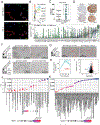
Figure 1.. Prioritization of DLK1 as a candidate immunotherapeutic target in neuroblastoma
(A) Schematic showing integration of mass spectrometry and RNA-sequencing to prioritize cell surface proteins as candidate immunotherapeutic targets. Proteins were filtered for surface expression, normal tissue expression and MS abundance. (B) Number of proteins identified in both cell lines and xenografts for each step of prioritization. (C) DLK1 RNA-sequencing data indicate high expression in a subset of neuroblastoma and normal tissue expression restricted to the adrenal/pituitary gland and ovary. (D) Enhancer rank plots of H2K27ac signal show a subset of neuroblastoma cell lines (N=6) have a super enhancer (red). X-axis represents the enhancer gene regions reported by LILY ordered by decreasing H3K27Ac signal (y-axis). The inflection point defines super enhancers. (E-G) Active super-enhancer correlates with higher DLK1 expression measured by RNA-sequencing, immunoblotting and immunohistochemistry of a neuroblastoma cell plug array. (H) Visualization of DLK1 locus highlighting H3K27Ac, H3K4me1 and H3K4me3 and CRC members in SK-N-Be(2)C. (I) High DLK1 mRNA expression is associated with poor event-free survival in high-risk neuroblastoma. See also Figures S1-S4.
Figure 2.
Figure 2.. DLK1 is a cell surface protein upregulated in neuroblastoma and plays a role in differentiation
(A) Immunofluorescence shows colocalization of DLK1 (green) with the cell surface marker Cadherin (red) in cell lines with high DLK expression. (B) Flow cytometry validates cell surface expression in a panel of cell lines: DLK1+ (NB: SK-N-Be(2)C and NBL-S), DLK1-Low (NB:NB-69) and negative (NB:NLF; SW620 and HEK293T). (C) Immunohistochemistry using tissue microarrays of xenograft neuroblastoma and pediatric normal tissues. (D) Representative neuroblastoma cores with high and low DLK1 expression. (E) Expression of DLK1 in Treehouse Childhood Cancer Initiative dataset. (F) SK-N-Be(2)C and SH-SY5Y neuroblastoma cells treated with all trans retinoic acid (ATRA) exhibit depleted expression of DLK1 and induced differentiation. Images of SK-N- Be(2)C and SH-SY5Y cells treated with DMSO alone and 5uM ATRA. (G) Western blotting demonstrating robust knockdown of DLK1 compared to control in SK-N-Be(2)C cells. (H) Incucyte NeuroTrack analysis shows differentiation phenotype (neurite length) following shRNA DLK1 knockdown. (I) Mass spectrometry analysis of control and shDLK1 cells (biological and technical duplicate) indicates DLK1 is the fourth most downregulated protein in the proteome. (J) DLK1 knockdown shows upregulation of NOTCH3 and downregulation of cell cycle, consistent with differentiation. See also Figures S5-S6.
Figure 3.
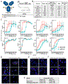
Figure 3.. Characterization of ADCT-701 shows binding, internalization and cytotoxicity
(A) ADCT-701 drug structure is comprised of valine/alanine linker with the PBD dimer SG3199. (B) Flow cytometry on a panel of cell lines shows DLK1 cell surface copy number and HuBA-1-3D binding as determined by EC50. Data are presented as mean antigen density and standard error of the mean (SEM). (C) IC50 on a panel of cell lines with variable DLK1 expression (mean+/−SD) (NBio=3; NTech=2). (D-E) Confocal microscopy shows colocalization of ADCT-701 with LAMP1 in A204 cells (DLK1+ cells) that is absent with the non-targeting control B12-PL1601. (F) Confocal microscopy shows no internalization of ADCT-701 in NCI-H1299 (DLK1-) cells. (G) Surface plasma resonance (SPR) shows comparable binding to human and cynomolgous monkey DLK1.
Figure 4.
Figure 4.. ADCT-701 shows specific and potent anti-tumor activity in neuroblastoma models expressing DLK1
(A) DLK1 RNA levels as measured by TPM. Red asterisks indicate models used for ADCT-701 efficacy studies. (B) DLK1 IHC staining of a xenograft array with variable DLK1 expression across models. (C) Tumor volumes (cm3) were measured for the saline control (red), non-targeting control ADC – B12-PL1601 (blue) and ADCT-701 (green). Arrows indicate the administration of a second dose (black) and tumor collection (red). Each model was scored (PD1/PD2-Progressive Disease; SD-Stable Disease; CR-Complete Response; MCR-Maintained Complete Response). (D) IHC of CDX/PDX array (top row) are compared to the relapse tumors (bottom row). See also Figure S7.

Update of

References

    1. Maris JM (2010). Recent advances in neuroblastoma. N Engl J Med 362, 2202–2211. 10.1056/NEJMra0804577. - DOI - PMC - PubMed
    1. Matthay KK, Maris JM, Schleiermacher G, Nakagawara A, Mackall CL, Diller L, and Weiss WA (2016). Neuroblastoma. Nat Rev Dis Primers 2, 16078. 10.1038/nrdp.2016.78. - DOI - PubMed
    1. Pugh TJ, Morozova O, Attiyeh EF, Asgharzadeh S, Wei JS, Auclair D, Carter SL, Cibulskis K, Hanna M, Kiezun A, et al. (2013). The genetic landscape of high-risk neuroblastoma. Nature genetics 45, 279–284. 10.1038/ng.2529. - DOI - PMC - PubMed
    1. Sausen M, Leary RJ, Jones S, Wu J, Reynolds CP, Liu X, Blackford A, Parmigiani G, Diaz LA Jr., Papadopoulos N, et al. (2013). Integrated genomic analyses identify ARID1A and ARID1B alterations in the childhood cancer neuroblastoma. Nature genetics 45, 12–17. 10.1038/ng.2493. - DOI - PMC - PubMed
    1. Cheung NK, Zhang J, Lu C, Parker M, Bahrami A, Tickoo SK, Heguy A, Pappo AS, Federico S, Dalton J, et al. (2012). Association of age at diagnosis and genetic mutations in patients with neuroblastoma. JAMA 307, 1062–1071. 10.1001/jama.2012.228. - DOI - PMC - PubMed

MeSH terms

LinkOut - more resources