. 2024 Dec;300(12):107916.
doi: 10.1016/j.jbc.2024.107916. Epub 2024 Oct 24.

Distinct chemical structures inhibit the CEMIP hyaluronidase and promote oligodendrocyte progenitor cell maturation

Affiliations

Distinct chemical structures inhibit the CEMIP hyaluronidase and promote oligodendrocyte progenitor cell maturation

Alec Peters et al. J Biol Chem. 2024 Dec.

Abstract

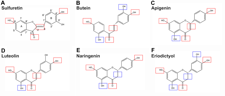
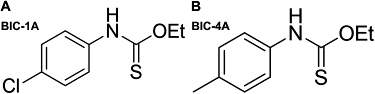
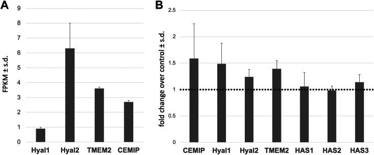
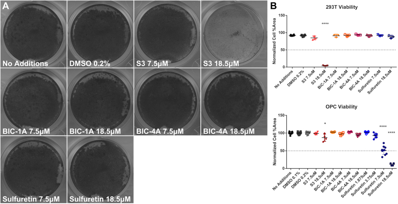
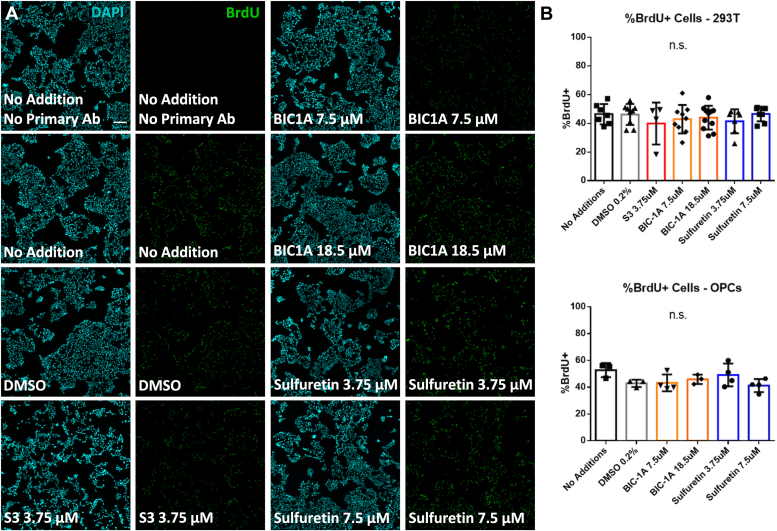
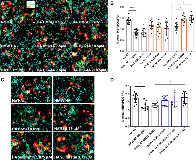
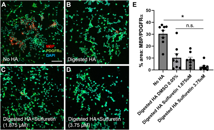
Growing evidence supports pathogenic roles for chronically elevated hyaluronidase activity in numerous conditions. Elevated expression of one such hyaluronidase, the Cell Migration Inducing and hyaluronan binding Protein (CEMIP), has been implicated in the pathogenesis and progression of several cancers as well as demyelinating diseases in the central nervous system (CNS). Developing effective and selective CEMIP inhibitors could therefore have efficacy in treating a variety of conditions where CEMIP is chronically elevated. Using two distinct screens for novel hyaluronidase inhibitors, we identified two synthetic thiocarbamates and one plant-derived flavonoid, sulfuretin, that effectively blocked CEMIP activity in live cells, including a tumorigenic cell line and primary cultures of oligodendrocyte progenitor cells (OPCs). None of these agents influenced cell proliferation, but they had differential dose-dependent and cell type-specific effects on cell survival. Furthermore, we found that each of these agents could promote oligodendrocyte maturation by OPCs in the presence of high molecular weight (>2 Mda) hyaluronan, the accumulation of which is linked to the inhibition of OPC maturation and remyelination failure in demyelinating diseases. These findings indicate that CEMIP can be inhibited through distinct chemical interactions and that CEMIP inhibitors have potential efficacy for treating demyelinating diseases or other conditions where CEMIP is elevated.

Keywords: CEMIP; flavonoids; hyaluronan; hyaluronidase; oligodendrocytes; sulfuretin.


Conflict of interest statement

Conflict of interest The authors declare that they have no conflicts of interest with the contents of this article.

Figures

Figure 1
Figure 1
Structures of flavonoids extracted from dahlia and coreopsis flowers. (A) Red boxes denote similarities between compound structures and sulfuretin, while blue boxes denote differences. The other structures are (B) butein, a chalcone; (C) apigenin, a flavone that has previously been used as a hyaluronidase inhibitor; (D) luteolin, another flavone; (E) naringenin, a compound in the flavonoid group of polyphenols; (F) eriodictyol, another flavone.
Figure 2
Figure 2
Chemical structures of thesynthetically-derivedthiocarbamates. (A) BIC-1A and (B) BIC-4A.
Figure 3
Figure 3
Novel synthetic flavonoids and sulfuretin inhibit CEMIP activity.A, Western blot showing CEMIP expression in 293T cells transfected with a CEMIP-expression vector. Actin was used as a loading control. B, representative HA agarose gel shows that 293T cells overexpressing CEMIP degrade HMW HA and that S3, BIC-1A, BIC-4A, and sulfuretin block this hyaluronidase activity in a dose-dependent manner. Note that the slight differences in the sizes of HMW HA in media alone compared to the vector control are within the range of normal HMW HA size variability. C, quantification of inhibitory activity of each compound in comparison to S3 in 293T cells. D, Western blot showing CEMIP expression in primary cultures of OPCs transfected with a CEMIP-expression vector. E, representative HA agarose gel showing that OPCs overexpressing CEMIP degrade HMW HA and that the hyaluronidase inhibitors block this activity. F, quantification of inhibitory activity of each compound in comparison to S3 in OPCs. ∗∗p < 0.01, ∗∗∗p < 0.001, ∗∗∗∗p < 0.0001, and n.s. – not significant.
Figure 4
Figure 4
Sulfuretin does not influence hyaluronidase expression in OPCs.A, relative expression (fragments per kilobase of transcript per million fragments mapped; FPKM) of hyaluronidase transcripts in mouse OPCs. Data were collected from the Brain RNA-Seq database (https://brainrnaseq.org). B, qPCR analysis of hyaluronidase and HA synthase transcripts in OPCs treated with 3.75 μM sulfuretin for 24 h compared to vehicle (DMSO)-treated controls (represented by the dashed line). There was no significant difference in expression of any of the genes analyzed.
Figure 5
Figure 5
Crystal violet viability assay of cells exposed to hyaluronidase inhibitors.A, representative images displaying 293T cells under normal growth conditions with indicated inhibitor doses. B, quantification of cell viability of 293T cells (upper) and OPCs (lower). ∗p < 0.05, and ∗∗∗∗p < 0.0001.
Figure 6
Figure 6
Cell proliferation is unaffected by hyaluronidase inhibitors.A, representative image of 5-bromo-2-deoxyuridine (BrdU) uptake of 293T cells exposed to indicated concentrations of hyaluronidase inhibitors. B, percentages of BrdU+ 293T cells (top panel) and OPCs (bottom panel) following treatment with inhibitors. Scale bar = 100 μm. n.s. – not significant.
Figure 7
Figure 7
Hyaluronidase inhibitors promote in vitro OPC maturation in the presence of HMW HA.A and C, representative images showing numbers of PDGFRα+ OPCs and MBP+ oligodendrocytes when exposed to HMW HA, or HMW HA with vehicle, synthetic inhibitors (A) or sulfuretin (B). B and D, quantification of OPC maturation in the presence of HMW HA, S3, and synthetically-derived hyaluronidase inhibitors. B, BIC-1A and BIC-4A both increase OPC maturation in the presence of HMW HA at 18.5 μM, while S3 is toxic to cells at this dose. D, sulfuretin is more potent than S3 at promoting OPC maturation when exposed to HMW HA. Scale bar = 30 μm. ∗p < 0.05, and ∗∗∗∗p < 0.0001.
Figure 8
Figure 8
Sulfuretin does not rescue the effects of digested HA on OPC maturation.AD, Representative immunohistochemical analyses of MBP (red) and PDGFRa (green) expression in primary mouse OPC cultures under differentiation conditions for 72 h and treated without HA (A), with HMW HA digested by CEMIP (B), (C) CEMIP-digested HA+ 1.875 μM sulfurtein, and (D) 3.75 μM sulfurein. Scale bar = 100 μm. E, quantification of immunohistochemical analyses in (AD). ∗p < 0.001, and n.s. = not significant.

Similar articles

References

    1. Walker C., Mojares E., Del Rio Hernandez A. Role of extracellular matrix in development and cancer progression. Int. J. Mol. Sci. 2018;19:3028. - PMC - PubMed
    1. Srivastava T., Sherman L.S., Back S.A. Dysregulation of hyaluronan homeostasis during white matter injury. Neurochem. Res. 2020;45:672–683. - PMC - PubMed
    1. De Luca C., Papa M. Matrix metalloproteinases, neural extracellular matrix, and central nervous system pathology. Prog. Mol. Biol. Transl. Sci. 2017;148:167–202. - PubMed
    1. Fawcett J.W. The extracellular matrix in plasticity and regeneration after CNS injury and neurodegenerative disease. Prog. Brain Res. 2015;218:213–226. - PubMed
    1. McAtee C.O., Barycki J.J., Simpson M.A. Emerging roles for hyaluronidase in cancer metastasis and therapy. Adv. Cancer Res. 2014;123:1–34. - PMC - PubMed

Publication types

MeSH terms

LinkOut - more resources